
Page 43 - GTM-3-3
P. 43
Global Translational Medicine Ocular changes in Alzheimer’s disease
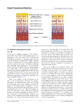
Figure 2. Schematic of the retina and the structural changes associated with Alzheimer’s disease. Created using BioRender.com.
3.3. Alzheimer’s pathology in the retina plaques were only detectable in the brain when mice
31
3.3.1. Aβ were 5 months old. Therefore, in this mouse model, Aβ
pathology in the retina occurred before it was present in
As discussed, a definitive diagnosis of AD requires a the brain. There is also evidence that retinal Aβ levels
31
post-mortem examination for pathological signs such as correlate with disease progression. The Aβ42 levels in the
Aβ plaques or NTs in the brain. The current literature post-mortem retina of individuals with MCI were 5 times
15
investigating the presence of Aβ plaques and NTs in the higher than in those with normal cognition. Moreover,
35
retina of AD subjects is summarized in Table 1. The first the Aβ42 levels in the AD subjects were significantly higher
evidence of Aβ deposits in the retina of patients with AD than in the MCI patients, revealing the accumulation of
was from a post-mortem study. Aβ plaques resembling Aβ42 with disease severity. 35
31
brain pathology were discovered in the post-mortem
retinas of definitive AD and MCI subjects, whereas plaques There is also evidence of a correlation between Aβ
were undetectable in the age-matched control group. burden in the eye and the brain. First, post-mortem retinal
31
Moreover, oral curcumin administration to label Aβ in live Aβ plaque levels, specifically in the superior temporal
AD patients followed by SLO has been used to reveal Aβ and inferior temporal regions of the retina, were closely
deposits in the retina. 22,32 Most deposits are found in the correlated to cerebral Aβ plaque levels. Furthermore,
35
inner layers and periphery of the retina, especially in the in vivo, retinal imaging on mouse models showed that
superior quadrant (Figure 3). 22,31,33 curcumin-labeled Aβ plaques in the retina correlated
23
There is also evidence of Aβ accumulation in the strongly to Aβ immunoreactivity in the cortex. These
retina in the pre-symptomatic AD stage. For example, results suggest that Aβ in the retina can estimate the degree
31
Aβ plaques were discovered post-mortem in the retina of Aβ pathology in the brain. Studies using retinal imaging
of an individual with normal cognition who had Aβ on post-mortem subjects also noted a similar morphology
22
plaques in their hippocampus. Investigation of murine between Aβ plaque in the brain and the retina. One
31
models has shown that retinal Aβ burden can be an early- difference is that plaques in the retina are 6 – 7 times smaller
22
stage biomarker for AD in vivo. These investigations use in diameter compared to those in the brain. Interestingly,
31
genetically engineered mouse models that overexpress the distribution of retinal Aβ plaques has been consistently
proteins with mutations identified in familial AD. 31,34,35 found surrounding vasculature. 22,31,36 In summary, these
The examination of APPSWE/PS1ΔE9 transgenic mice findings suggest that retinal Aβ burden reflects pathology
using curcumin Aβ labeling revealed that Aβ plaques were in the brain and therefore could be useful in the diagnosis
detectable in the retina of 2.5-month-old mice whereas of AD in living patients.
Volume 3 Issue 3 (2024) 4 doi: 10.36922/gtm.4094