
Page 129 - GTM-4-1
P. 129
Global Translational Medicine AA amyloidosis in rheumatoid arthritis
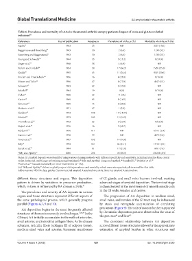
Table 6. Prevalence and mortality of sAAa in rheumatoid arthritis autopsy patients: Impact of sAAa and giAAa on lethal
outcomes 42
References Year of publication Autopsy, n Prevalence of sAAa, n (%) Mortality of sAAa, n/N (%)
Bayles 53 1943 23 ND 3/23 (13.0)
Baggenstoss and Rosenberg 54 1943 30 2 (6.6) 1/30 (3.3)
Rosenberg and Baggenstoss5 5 1943 30 2 (6.6) 1/30 (3.3)
Young and Schwedel 56 1944 33 5 (15.2) 0/33 (0)
Unger et al. 57 1948 58 4 (6.9) ND
Teilum and Lindahl 58 1954 28 17 (60.7) 7v28 (25.0)
Gedda 59 1955 45 11 (24.4) 9/45 (20.0)
Sinclair and Cruickshank 60 1956 16 4 (25.0) 0/16 (0)
Missen and Tailor 61 1956 47 8 (17.0) 4/47 (8.5)
Lebowitz 62 1963 62 6 (10.0) ND
Sokoloff 63 1964 19 0 (0) 0/19 (0)
Cohen 4 1968 42 11 (26) ND
Karten 64 1969 95 1 (1.05) ND
Gritsman 65 1969 15 6 (40.0) ND
Ozdemir et al. 66 1971 47 1 (2.1) ND
Gardner 67 1972 142 17 (11.97) ND
Püschel 68 1973 143 15 (10.5) ND
Vroninks et al. 69 1973 62 3 (4.84) 0/62 (0)
Hajzok et al. 70 1976 16 7 (43.7) ND
Eulderink 71 1976 111 ND 6/111 (5.4)
Rainer et al. 72 1978 79 ND 4/79 (5.0)
a Boers et al. 73 1987 132 14 (10.6) ND
Bély 74 1993 161 34 (21.1) 17/161 (11)
Suzuki et al. 75 1994 81 17 (21.0) 6/81 (7.4)
b Bély and Apáthy 76 2006 234 48 (20.5) 20/234 (8.5)
Notes: (i) Amyloid deposits were identified using various staining methods with different specificity and sensitivity, including toluidine blue, crystal
violet, Syrius red, and Congo red staining using Romhányi’s, Bély and Apáthy’s Congo red method, Bennhold’s, Puchtler et al., 78
45
46
77
a Boers et al. focused exclusively on renal involvement (n=132).
73
(iii) Bély and Apáthy did not explicitly report sAAa prevalence and mortality, which were retrospectively determined for this table.
b
76
Abbreviations: ND: No data; giAAa: Gastrointestinal amyloid A amyloidosis; sAAa: Systemic amyloid A amyloidosis.
different tissue structures and organs. This deposition of GI glands, and small veins become involved, marking
pattern is driven by variations in precursor production, advanced stages of amyloid deposition. The terminal stage
which, in turn, is influenced by RA disease activity. 41 is characterized by the involvement of smooth muscle cells
The prevalence and severity of AA deposits in various in the GI walls, venules, and nerves.
organs and tissue structures represent different aspects of The progression of AA deposition in medium-sized,
the same pathological process, which generally progress small veins, and venules of the GI tract may be influenced
parallel (Figures 6,7, 9 and 10). by stasis and retrograde accumulation of circulating
precursors (Figure 9). The role of stasis is further supported
AA deposition begins in the most frequently affected
structures of the most commonly involved organ. 3,74,79 In the by the similar deposition patterns observed in the veins of
43
41
GI tract, AA initially accumulates in the walls of arterioles, the pancreas and liver
small arteries, and interstitial collagen fibers. As the disease The consistent relationship between AA deposition
advances, reticulin fibers (collagen III of adipose tissue), across different tissue structures allows for the approximate
medium-sized veins and arteries, basement membranes estimation of amyloid burden in other structures and
Volume 4 Issue 1 (2025) 121 doi: 10.36922/gtm.5325