
Page 114 - GTM-4-2
P. 114
Global Translational Medicine Platelet aggregation inhibition by 2-isoxazolines
[ddd, J = 1.7, 1.4, 0.5 Hz]), 7.45 (1H, ddd, J = 8.1, 7.7, of labeled antibodies: CD41a-fluorescein isothiocyanate
0.5 Hz), and 7.64 (1H, ddd, J = 7.7, 1.7, 1.2 Hz). C (FITC) and CD61-phycoerythrin (PE). Further analysis
13
NMR: δ 41.8 (1C, s), 52.2 (1C, s), 77.8 (1C, s), 115.0 – of platelet surface markers GPIIa (CD41a) and GPIIIb
115.1 (2C, 115.0 (s), 115.1 (s)), 127.3 (1C, s), 130.2 (1C, (CD61) was performed using a flow cytometer Perlong
s), 132.1 (1C, s), 155.7 (1C, s), 161.2 (1C, s), and FC2060 (Perlong Medical Equipment, China). The
170.2 (1C, s). gating strategy was based on determining the number of
+
+
(vi) Compound 6: Methyl ester of 3-(4-fluorophenyl)-2- double positive (CD41a CD61 ) cells on the cytogram.
isoxazoline carboxylic acid. The compound has a yield Methyl ester of (+)-(S)-alpha-(o-chlorophenyl)-
[3
of 84% and a melting point between 171 and 173°С. 6,7-dihydrothieno .2-c]pyridine-5(4H)-acetic acid
The IR spectrum (KBr, cm ) shows the following (Clopidogrel), which is currently used as an antiplatelet
-1
peaks: 3101, 3092 (C-H aromatic), 1806, 1263 (COO), agent (final concentration 5 and 10 mmol/L, respectively)
1651 (C = N), and 1603, 1507 (C = C aromatic). The was used as a positive control.
UV spectrum (EtOH, λ max , nm) is 271. The H NMR 2.4. Statistical analysis
1
shows the following: δ 2.94 (2H, dd, J = 15.5, 7.4 Hz),
3.75 (3H, s), 5.27 (1H, dd, J = 7.9, 6.8 Hz), 7.04 (2H, To ensure the reliability of the results, all measurements
ddd, J = 8.7, 1.0, 0.6 Hz), and 7.93 (2H, ddd, J = 8.7, 1.6, were performed in triplicate, which eliminated random
13
0.6 Hz). The C NMR shows δ 41.8 (1C, s), 52.2 (1C, s), errors and increased the reliability of the data. The platelet
77.8 (1C, s), 115.4 (2C, s), 125.3 (1C, s), 128.6 (2C, s), aggregation results were determined as the maximum
155.7 (1C, s), 162.5 (1C, s), and 170.2 (1C, s). amplitude expressed as a percentage. This indicator
was calculated using specialized software (version 2.1,
2.2. Light transmission method MedCalc) that analyzed the data obtained during the
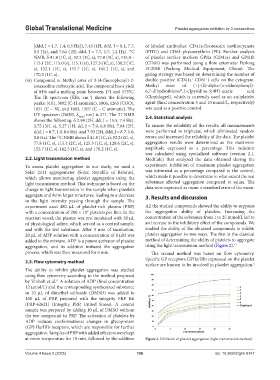
To assess platelet aggregation in our study, we used a experiment. Inhibition of maximum platelet aggregation
Solar 2111 aggregometer (Solar, Republic of Belarus), was estimated as a percentage compared to the control,
which allows monitoring platelet aggregation using the which made it possible to determine to what extent the test
light transmission method. This technique is based on the substance affected aggregation compared to saline. The
change in light transmission in the sample when platelets data were expressed as mean ± standard error of the mean.
aggregate and form larger structures, leading to a decrease 3. Results and discussion
in the light intensity passing through the sample. The
experiment used 480 μL of platelet-rich plasma (PRP) All the studied compounds showed the ability to suppress
with a concentration of 200 × 10 platelets per liter. In the the aggregation ability of platelets. Increasing the
9
reaction vessel, the plasma was pre-incubated with 20 μL concentration of the substance from 1 to 25 mmol/L led to
of physiological saline, which served as a control sample, an increase in the inhibitory effect of the compounds. We
and with the test substance. After 3 min of incubation, studied the ability of the obtained compounds to inhibit
20 μL of ADP solution with a concentration of 8 μM was platelet aggregation in two ways. The first is the classical
added to the mixture. ADP is a potent activator of platelet method of determining the ability of platelets to aggregate
aggregation, and its addition initiated the aggregation using the light transmission method (Figure 2). 11
process, which was then measured for 6 min. The second method was based on flow cytometry.
Specific GP receptors GPIIa/IIIb expressed on the platelet
2.3. Flow cytometry method
surface are known to be involved in platelet aggregation.
5
The ability to inhibit platelet aggregation was studied
using flow cytometry according to the method proposed
by Vinholt et al. A solution of ADP (final concentration
11
12 μmol/L) and the corresponding synthesized substance
in 10 μL of dimethyl sulfoxide (DMSO) was added to
100 μL of PRP, prepared with the integrity PRP Kit
(PRP-62621) (Integrity PRP, United States). A control
sample was prepared by adding 10 μL of DMSO without
the test compound to PRP. The activation of platelets by
ADP induces conformational changes in glycoprotein
(GP) IIa/IIIb receptors, which are responsible for further
aggregation. Samples of PRP with added effectors were kept
at room temperature for 15 min, followed by the addition Figure 2. Inhibition of platelet aggregation (light transmission method)
Volume 4 Issue 2 (2025) 106 doi: 10.36922/gtm.8147