
Page 121 - IJB-10-1
P. 121
International Journal of Bioprinting Droplet-based bioprinting of tumor spheroids
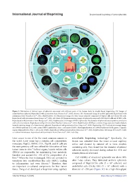
Figure 3. Fabrication of distinct types of spheroids associated with different parts of the human body by droplet-based bioprinting. (A) Images of
neuroblastoma spheroid. Reproduced with permission from Utama et al. , 2020, iScience. (B) Fluorescent image of an REC spheroid. Reproduced with
74
permission from Trondle et al. , 2021, Biofabrication. (C) Fluorescent image of a liver tumor spheroid composed of HepG2 cells and EA.hy 926 cells.
76
Reproduced with permission from Hong et al. , 2021, Adv Mater. (D) Immunostaining images of spheroids composed of 60 HepG2 cells and 40 HSC cells.
80
Reproduced with permission from Zhang et al. , 2022, Biofabrication. (E) Images of hESCs spheroids. The diameter of spheroids was in positive correlation
81
with cell concentration. Reproduced with permission from Faulkner-Jones et al. , 2013, Biofabrication. (F) Phase-contrast image and calcein and ethidium
42
bromide staining image of a breast tumor spheroid. Reproduced with permission from Ling et al. , 2015, Engineering. (G) Confocal microscopy image
86
of a SW620 spheroid (G-I), red = actin (TRITC-phalloidin), blue = cell nuclei (DAPI). Histological section of a SW620 spheroid (G-II), green = hypoxic
region (HypoxyProbe), blue = cell nuclei (DAPI). Reproduced with permission from Johnson et al. , 2022, Biofabrication. (H) Image of (CAL27)–CAFs
87
co-culture microtissues. Reproduced with permission from Chen et al. , 2021, Lab Chip.
46
Liver cancer is one of the five most common cancers in microfluidic bioprinting technology. Specifically, the
80
the world. Liver tissue has a complex cell composition; bioink was extruded from the inner co-axial capillary
nowadays, HepG2, SMMC-7721, Hep3B, and J5 cells are orifice and sheared by mineral oil to form droplets
the main primary cell lines utilized for fabrication of liver containing cells. They found that the diameter of printed
77
tumor tissue in vitro. In liver organs, hepatic stellate cells spheroids rapidly decreased during culture for 10 h and
(HSCs) are responsible for maintaining liver metabolic remain plateaued afterward.
function and account for ~8% of the composition of a
liver. When the liver is damaged, HSCs are activated to Cell viability of structured spheroids was above 90%
78
transform into myofibroblast-like cells (MFC), leading after 7-day culture. They fabricated uniform spheroids
7
to inflammation and even fibrosis. Therefore, they composed of HepG2/C3A cells (5 × 10 cells/ml) and
79
7
are important co-cultured components for liver tumor endothelial cells (EA.hy 926) (5 × 10 cells/ml) with a
tissue. Hong et al. developed a bioprinter using capillary diameter of ~250 μm (Figure 3C) in a high-throughput
Volume 10 Issue 1 (2024) 113 https://doi.org/10.36922/ijb.1214