
Page 125 - IJB-10-1
P. 125
International Journal of Bioprinting Droplet-based bioprinting of tumor spheroids
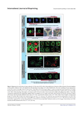
Figure 4. Representative applications of tumor spheroids. (A) The ratio of HSCs affects the morphology and function of liver spheroids. Presented numbers
are the number of HepG2 + HSC cells. Reproduced with permission from Zhang et al. , 2022, Biofabrication. (B) In vitro modeling of tumor angiogenesis.
81
Some GSCs moved to the edge of vascular channel after 7-day culture (left). Microscope images of Y-patterns containing lumen formed from ECs (right).
Reproduced with permission from Lee et al. , 2015, Proceedings of the 41st Annual Northeast Biomedical Engineering Conference (left) and from Trondle
96
et al. , 2019, J Tissue Eng Regen Med (right). (C) Invasion model of (CAL27)–CAFs. CAL27 tumor cells invaded surrounding tissues within 7 days (left),
97
and invasion phenomenon was significantly inhibited with the treatment of 5-FU (right). The double arrow indicates the direction and trace of tumor
cell invasion. Reproduced with permission from Chen et al. , 2021, Lab Chip (left) and from Chen et al. , 2022, J Mater Chem B (right). (D) Fluorescent
46
101
images of live/dead cells of brain tumor spheroid before and after PTT treatment. NSC spheroids were pretreated by photosensitive rGO-BPEI-PEG
nanocomposites and exposed to the near-infrared (NIR) laser irradiation. Reproduced with permission from Lee et al. , 2020, Microsyst Nanoeng. (E)
103
Doxorubicin suppressed the survival and proliferation of LCC6/Her2 tumor spheroids. Presented numbers are the dosage of doxorubicin (unit: nM).
104
Reproduced with permission from Yu et al. , 2010, Lab Chip.
Volume 10 Issue 1 (2024) 117 https://doi.org/10.36922/ijb.1214