
Page 122 - IJB-10-1
P. 122
International Journal of Bioprinting Droplet-based bioprinting of tumor spheroids
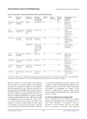
Table 2. Representative tumor spheroids fabricated by droplet-based bioprinting
Tumor Fabrication Printing ma- Cell types, Viability Culture Spheroid Morphology and Ref.
method trixes (% w/v) (concentration (%) time (days) diameter physiological
[× 10 cells/ml]) (μm) responses
7
Neuroblastic Microvalve-based Alginate SK-N-BE(2) (25) >95 3 ~200 Expression of 74
tumor bioprinting Ki67 protein,
HIF1α-positive
cells, cell ratio,
and distribution
REC Piezoelectric ink- Matrigel-colla- iREC (0.5–2.5) ~92 7 ~50 Spheroid 76
spheroid jet bioprinting gen I, Fibrin- diameter, inner
ogen structure, kid-
ney-specific gene
signatures
Liver tumor Microfluidic Neural collagen HepG2 (5) + >90 7 ~250 Expression of 80
bioprinting solution (4%) EA.hy926 (5) MRP2, albumin,
and CD31, blood
vessels
DMEM (90%) HepG2 + HSC ~98 6 100 Type I collagen, 81
(a total number spheroid size, and
of 100, the ratio morphology
of HSCs ranges
from 0% to
100%)
hESC Microvalve-based hESC SFM hESCs (0.3) >89 3 250–600 Spheroid mor- 42
spheroid bioprinting phology, Oct-4
pluripotency
marker
Breast tumor Microvalve-based Gelatin (3%) MCF-7 (0.1) >95 7 200 Spheroid mor- 86
bioprinting phology, size, and
compactness
Colorectal Microvalve-based Gelatin (1%)– SW620 (0.015) ~80 7 ~200 Spheroid 87
tumor bioprinting alginate (10%) morphology,
compactness, and
inner structure
Oral tumor Acoustic bioprint- Gelatin (5%) CAL27 (10) + >94 5 ~100 Cell distribution 46
ing CAFs (10) in microenviron-
ment
Abbreviations: DMEM, Dulbecco’s Modified Eagle Medium; hESC, human embryonic stem cell; HIF1α, hypoxia-inducible factor 1-alpha; MRP2, multi-
drug resistance-associated protein 2; Oct-4, octamer-binding transcription factor 4; SFM, serum- and feeder-free medium.
manner. Compared to non-structured renal spheroid, at over 98%. By measuring the size and secretion of type
the EA.hy 926 cells covered the surface of the structured I collagen of finally formed multicellular spheroids, they
spheroid and maintained the stability of morphology of demonstrated that the initial ratio of HSCs and HepG2
the liver spheroid for 10 days. They also found that the cells affected the morphology and function of these
gene expression of multidrug resistance-associated protein spheroids. Compared with common random loading
2 (MRP2), albumin, and CD31 was upregulated in the co- process, single-cell printing produced liver spheroids that
cultures. To match the complexity of liver tumors in vivo, were more uniform.
Zhang et al. utilized microfluidic flow cytometric printing
technology to print HepG2 and HSC cells into microwells 3.4. Human embryonic stem cell spheroids
for multicellular liver spheroid fabrication (Figure 3D). Human embryonic stem cells (hESCs) can proliferate
81
As a cell-by-cell fabrication method, microfluidic flow infinitely and differentiate into any functional cells in
cytometric printing technology allows for the control vitro. hESC spheroids can be utilized to study the
82
of cell numbers and types for each spheroid. Using this formation of tumors. However, hESCs are very fragile
method, they found that cell viability could be maintained and can spontaneously differentiate when stimulated
Volume 10 Issue 1 (2024) 114 https://doi.org/10.36922/ijb.1214