
Page 140 - IJB-10-1
P. 140
International Journal of Bioprinting Bioprinting organoids for toxicity testing
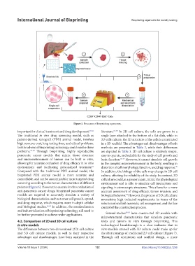
Figure 5. Processes of bioprinting a pancreas.
important for clinical treatment and drug development. 53-56 literature. 69-73 In 2D cell culture, the cells are grown in a
The traditional in vivo drug screening model, such as single layer attached to the bottom of a flat dish, while in
patient-derived xenograft (PDX) animal model, involves 3D cells culture, the 3D structure of the cells is constructed
high resource cost, long testing time, and ethical problems, in a 3D scaffold. The advantages and disadvantages of both
but the advent of bioprinting technology could resolve these methods are presented in Table 2, while their differences
problems. 57-60 Through bioprinting, highly reproducible are depicted in Table 3. 2D cell culture is relatively simple,
pancreatic cancer models that mimic tissue structure easy to operate, and suitable for the study of cell growth and
and microenvironment of human can be built in vitro, basic function. 74-77 However, it cannot simulate cell growth
allowing for accurate evaluation of drug efficacy in in vitro in the complex microenvironment in the body, resulting in
environment and facilitating personalized treatment distortion of cell morphology, function, and drug response.
78
61
Compared with the traditional PDX animal model, the In addition, the biology of the cells may change in 2D cell
bioprinted PDX animal model is more accurate and culture, affecting the reliability of the study. In contrast, 3D
controllable, and can be customized for more targeted drug cell culture could, at a greater extent, mimic the physiological
screening according to the tumor characteristics of different environment and is able to simulate cell interactions and
patients (Figure 6). However, to execute in vitro evaluation of signaling in stereoscopic structures. This allows for a more
anti-pancreatic cancer drugs, bioprinted pancreatic cancer accurate assessment of drug efficacy, tumor invasion, and
models are required to accurately simulate a variety of biological behavior. However, the process of 3D cell culture
79
biological characteristics, such as tumor cell growth, spread, necessitates high technical requirements, in terms of the
and drug response, which requires more in-depth cellular selection of scaffold materials, cell arrangement, and the fine
and biological studies. 62-69 In addition, the standardization control of the construction process.
and industrialization of bioprinting technology still need to 80-83
be further promoted to achieve wider applications. Several studies have constructed 3D models with
microstructural characteristics that simulate pancreatic
4.2. Comparison of 2D and 3D cell culture islets and tumors in vitro through bioprinting. This
in vitro models technological breakthrough is a clear indicator that in
The differences between two-dimensional (2D) cell culture vitro models created with 3D culture could make up for
and 3D cell culture models, as well as their respective the shortcomings of traditional 2D cell culture (Figure 7).
advantages and disadvantages, have been analyzed in the Through cell orientation and scaffold design, a more
Volume 10 Issue 1 (2024) 132 https://doi.org/10.36922/ijb.1256