
Page 37 - IJB-10-1
P. 37
International Journal of Bioprinting Bioprinted organ-on-a-chip with biomaterials
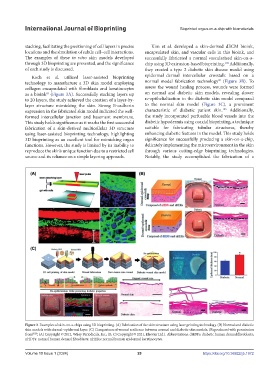
stacking, facilitating the positioning of cell layers in precise Kim et al. developed a skin-derived dECM bioink,
locations and the simulation of subtle cell–cell interactions. encapsulated skin, and vascular cells in this bioink, and
The examples of these in vitro skin models developed successfully fabricated a normal vascularized skin-on-a-
through 3D bioprinting are presented, and the significance chip using 3D extrusion-based bioprinting. Additionally,
104
of each study is discussed. they created a type 2 diabetic skin disease model using
Koch et al. utilized laser-assisted bioprinting epidermal–dermal intercellular crosstalk based on a
90
technology to manufacture a 3D skin model employing normal model fabrication technology (Figure 3B). To
collagen encapsulated with fibroblasts and keratinocytes assess the wound healing process, wounds were formed
as a bioink (Figure 3A). Successfully stacking layers up on normal and diabetic skin models, revealing slower
85
to 20 layers, the study achieved the creation of a layer-by- re-epithelialization in the diabetic skin model compared
layer structure mimicking the skin. Strong E-cadherin to the normal skin model (Figure 3C), a prominent
105
expression in the fabricated skin model indicated the well- characteristic of diabetic patient skin. Additionally,
formed intercellular junction and basement membrane. the study incorporated perfusable blood vessels into the
This study holds significance as it marks the first successful diabetic hypodermis using coaxial bioprinting, a technique
fabrication of a skin-derived multicellular 3D structure suitable for fabricating tubular structures, thereby
using laser-assisted bioprinting technology, highlighting enhancing diabetic features in the model. This study holds
3D bioprinting as an excellent tool for mimicking organ significance for successfully producing a skin-on-a-chip,
functions. However, the study is limited by its inability to delicately implementing the microenvironment in the skin
reproduce the skin’s unique function due to a restricted cell through various cutting-edge bioprinting technologies.
source and its reliance on a simple layering approach. Notably, the study accomplished the fabrication of a
Figure 3. Examples of skin-on-a-chips using 3D bioprinting. (A) Fabrication of the skin structure using laser printing technology. (B) Normal and diabetic
skin models with dermal–epidermal layer. (C) Comparison of wound resilience between normal and diabetic skin models. (Reproduced with permission
from 85,90 ; (A) Copyright © 2012, Wiley Periodicals, Inc.; (B, C) Copyright © 2021, Elsevier Ltd.). Abbreviations: dHDFs: diabetic human dermal fibroblasts;
nHDFs: normal human dermal fibroblasts; nHEKs: normal human epidermal keratinocytes.
Volume 10 Issue 1 (2024) 29 https://doi.org/10.36922/ijb.1972