
Page 42 - IJB-10-1
P. 42
International Journal of Bioprinting Bioprinted organ-on-a-chip with biomaterials
research. Owing to the absence of high-performance emulate the complex physiological structure of the liver
130
in vitro kidney disease models, the development of such and regulate its functionality. However, in vitro models
139
models, considering the abovementioned examples, holds created using traditional methods face challenges in
significant promise for advancing new treatments or drug integrating a multilayer structure comprising different liver
tests for kidney diseases. 132 cell types into a unified platform. Recently, to establish
140
a liver-specific microenvironment, liver-derived dECM
3.4. Liver (LdECM) has emerged as a bioink for 3D bioprinting
The liver, one of the body’s largest organs, plays a crucial role applications. In contrast to conventional manufacturing
27
in governing the body’s overall metabolism, encompassing methods, 3D bioprinting enables precise cell placement and
the regulation of blood sugar levels and the processing can replicate cell–cell interactions by constructing intricate
of various bodily substances. Furthermore, it actively liver structures within a single platform. Consequently,
133
20
contributes to the circulation and management of vital 3D bioprinting models can faithfully recreate the
nutrients such as carbohydrates, fats, hormones, vitamins, intricate 3D architecture and microenvironment of liver
and minerals while serving as a detoxification hub for tissue, closely mirroring actual liver pathophysiology
harmful compounds. The array of recognized liver and enhancing the realism and practicality of research
134
disorders is extensive, encompassing conditions such as findings. This technology can significantly contribute to a
hepatitis, liver cancer, and fatty liver disease. The ability deeper understanding of the mechanisms underlying the
135
to replicate diverse liver functions in vitro holds significant development and progression of liver diseases, as well as the
implications for tissue engineering, liver regenerative development of treatment and prevention strategies. The
medicine, and advancements in drug development. 136 value of this approach is exemplified by in vitro liver models
To establish an effective 3D in vitro model of the generated via 3D bioprinting with the LdECM bioink.
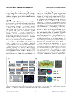
liver, it is crucial to recreate the distinctive sinusoidal Lee et al. pioneered the development of an LdECM
structure of the liver while ensuring functional maturity. bioink, incorporating various hepatic cells, and utilized
137
Within these sinusoids, various cell types, including liver 3D extrusion-based bioprinting to create an initial in vitro
sinusoidal endothelial cells, hepatocytes, hepatic stellate liver model. Building upon this technology for normal
27
cells, and Kupffer cells, are positioned within the 3D ECM liver model construction, they established an in vitro
microenvironment, forming two fluidic channels in the liver fibrosis model by encapsulating activated stellate
sinusoids. Several microfabrication methods, such as cells, known inducers of liver fibrosis, within gelatin
138
photolithography and etching, have been employed to (Figure 6A). This innovative approach incorporated most
Figure 6. Examples of liver-on-a-chip and placenta-on-a-chip fabricated via 3D bioprinting. (A) Liver-on-a-chip production process and functions. (B)
Placenta-on-a-chip design and cell location by date (Reproduced with permission from 27,157 ; (A) Copyright © 2020, American Chemical Society; (B)
Copyright © 2016, American Chemical Society). Abbreviations: EGF: epidermal growth factor; FN: fibronectin.
Volume 10 Issue 1 (2024) 34 https://doi.org/10.36922/ijb.1972