
Page 39 - IJB-10-1
P. 39
International Journal of Bioprinting Bioprinted organ-on-a-chip with biomaterials
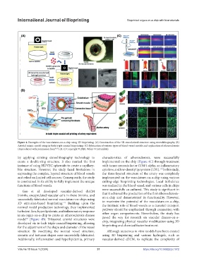
Figure 4. Examples of the vasculatures-on-a-chip using 3D bioprinting. (A) Construction of the 3D-vascularized structure using stereolithography. (B)
Arterial mimic model using in-bath triple coaxial bioprinting. (C) Fabrication of various types of blood vessel models and application of atherosclerosis
(Reproduced with permission from 91,92 ; (B, C) Copyright © 2020, Wiley-VCH GmbH).
by applying existing stereolithography technology to characteristics of atherosclerosis, were successfully
create a double-ring structure. It also marked the first implemented on the chip (Figure 4C) through treatment
instance of using HUVEC spheroids to create a capillary- with tumor necrosis factor (TNF)-alpha, an inflammatory
like structure. However, the study faced limitations in cytokine, and low-density lipoprotein (LDL). In this study,
118
expressing the complex, layered structure of blood vessels the three-layered structure of the artery was completely
and relied on limited cell sources. Consequently, the study implemented on the vasculature-on-a-chip using various
is constrained in its ability to fully implement the unique cutting-edge bioprinting technologies. Local turbulence
functions of blood vessels. was realized in the blood vessel, and various cells in chips
were successfully co-cultured. This study is significant in
Gao et al. developed vascular-derived dECM
bioinks, encapsulated vascular cells in these bioinks, and that it achieved the production of the first atherosclerosis-
on-a-chip and demonstrated its functionality. However,
successfully fabricated normal vasculature-on-chips using to maximize the potential of the vasculature-on-a-chip,
3D extrusion-based bioprinting. Building upon the the intrinsic role of blood vessels as a material transport
117
normal model production technology, they implemented pathway should be emphasized through connection with
turbulent flow, hyperlipidemia, and inflammatory response other organ compartments. Nevertheless, the study has
in an organ-on-a-chip to create an atherosclerosis disease paved the way for research on vascular disease-on-a-
model (Figure 4B). Trilayered arterial structures were chip, integrating physical vascular modification using 3D
92
developed via in-bath triple-coaxial bioprinting, allowing bioprinting and chemical factor treatment.
for the adjustment of the shape and diameter of the vessel
structure. By modifying the normal vessel structure, Although numerous in vitro models have been created
stenotic and tortuous shapes were successfully fabricated. using 3D bioprinting and various hydrogels, such as
Additionally, inflammation and hyperlipidemia, primary vascular-derived dECM, to replicate the complexity of
Volume 10 Issue 1 (2024) 31 https://doi.org/10.36922/ijb.1972