
Page 41 - IJB-10-1
P. 41
International Journal of Bioprinting Bioprinted organ-on-a-chip with biomaterials
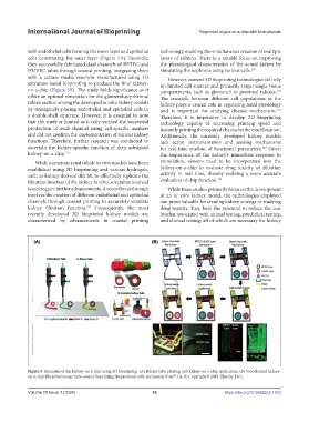
with endothelial cells forming the inner layer and epithelial technology, enabling the simultaneous creation of multiple
cells constituting the outer layer (Figure 5A). Secondly, layers of kidneys. There is a notable focus on improving
they successfully fabricated dual channels of RPTEC and the physiological characteristics of the actual kidney by
HUVEC tubes through coaxial printing, integrating them simulating the nephrons using various cells. 130
with a culture media reservoir manufactured using 3D However, current 3D bioprinting technologies still rely
extrusion-based bioprinting to produce the final kidney- on limited cell sources and primarily target single tissue
on-a-chip (Figure 5B). The study holds significance as it compartments, such as glomeruli or proximal tubules.
129
offers an optimal simulation for the glomerulus/proximal The crosstalk between different cell populations in the
tubule section among the developed in vitro kidney models kidney plays a crucial role in regulating renal physiology
by strategically placing endothelial and epithelial cells in and is important for studying disease mechanisms.
131
a double-shell structure. However, it is essential to note Therefore, it is imperative to develop 3D bioprinting
that the study is limited as it only verified the successful technology capable of increasing printing speed and
production of each channel using cell-specific markers instantly printing the required channel at the exact location.
and did not confirm the implementation of various kidney Additionally, the currently developed kidney models
functions. Therefore, further research was conducted to lack active instrumentation and sensing mechanisms
ascertain the kidney-specific function of their advanced for real-time readout of functional parameters. Given
129
kidney-on-a-chip. 125 the importance of the kidney’s immediate response to
While numerous renal tubule in vitro models have been stimulation, sensors need to be incorporated into the
established using 3D bioprinting and various hydrogels, kidney-on-a-chip to evaluate drug toxicity or filtration
such as kidney-derived dECM, to effectively replicate the activity in real time, thereby enabling a more accurate
131
filtration function of the kidney in vitro, several unresolved evaluation of chip function.
issues require further advancements. A recent breakthrough While these studies primarily focus on the development
involves the creation of different endothelial and epithelial of an in vitro kidney model, the technologies employed
channels through coaxial printing to accurately simulate can prove valuable for creating kidney analogs or studying
kidney filtration function. Consequently, the most drug toxicity. They have the potential to reduce the cost
129
recently developed 3D bioprinted kidney models are burden associated with animal testing, preclinical testing,
characterized by advancements in coaxial printing and clinical testing, all of which are necessary for kidney
Figure 5. Examples of the kidney-on-a-chip using 3D bioprinting. (A) Bilayer tube printing and kidney-on-a-chip application. (B) Vascularized kidney-
on-a-chip fabricated using triple coaxial bioprinting (Reproduced with permission from ; (A, B) Copyright © 2019, Elsevier Ltd.).
94
Volume 10 Issue 1 (2024) 33 https://doi.org/10.36922/ijb.1972