
Page 371 - IJB-10-1
P. 371
International Journal of Bioprinting Transdermal vitamin C delivery by MNPs
3.3. Investigation of puncture performance and MNPs. Our results suggested that MNPs could be used for
skin healing drug delivery in a minimally invasive manner.
Three-arm MNPs were selected to evaluate the puncture
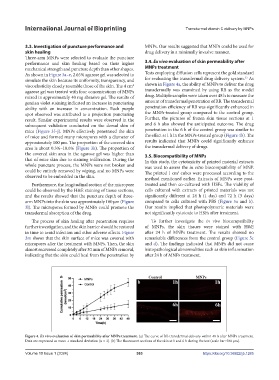
performance and skin healing based on their higher 3.4. Ex vivo evaluation of skin permeability after
mechanical strength and puncture depth than other shapes. MNPs treatment
As shown in Figure 3a–e, 2.65% agarose gel was selected to Tests employing diffusion cells represent the gold standard
33
simulate the skin because its uniformity, transparency, and for evaluating the transdermal drug delivery system. As
viscoelasticity closely resemble those of the skin. The 4 cm shown in Figure 4a, the ability of MNPs to deliver the drug
2
agarose gel was treated with four concentrations of MNPs transdermally was examined by using RB as the model
mixed in approximately 40 mg abrasive gel. The results of drug. Multiple samples were taken over 48 h to measure the
gentian violet staining indicated an increase in puncturing amount of transdermal penetration of RB. The transdermal
ability with an increase in concentration. Each purple penetration efficiency of RB was significantly enhanced in
spot observed was attributed to a projection puncturing the MNPs-treated group compared to the control group.
result. Similar experimental results were observed in the Further, the pictures of frozen skin tissue sections at 1
subsequent validation conducted on the dorsal skin of and 6 h also showed the anticipated outcome. The drug
mice (Figure 3f–j). MNPs effectively penetrated the skin penetration in the 6 h of the control group was similar to
of mice and formed many micropores with a diameter of the effect at 1 h in the MNPs-treated group (Figure 4b). The
approximately 100 μm. The proportion of the covered skin results indicated that MNPs could significantly enhance
area is about 0.3%–10.6% (Figure 3k). The proportion of the transdermal delivery of drugs.
the covered skin area in the agarose gel was higher than 3.5. Biocompatibility of MNPs
that of mice skin due to staining infiltration. During the In this study, the cytotoxicity of printed material extracts
whole puncture process, the MNPs were not broken and was used to assess the in vitro biocompatibility of MNP.
could be entirely removed by wiping, and no MNPs were The printed 1 cm cubes were processed according to the
3
observed to be embedded in the skin. method mentioned earlier. Extracts of MNPs were post-
Furthermore, the longitudinal section of the micropore treated and then co-cultured with HSFs. The viability of
could be observed by the H&E staining of tissue sections, cells cultured with extracts of printed materials was not
and the results showed that the puncture depth of three- significantly different at 24 h (1 day) and 72 h (3 days)
arm MNPs into the skin was approximately 100 μm (Figure compared to cells cultured with PBS (Figure 5a and b).
3l). The micropores formed by MNPs could promote the Our results implied that photopolymeric materials were
transdermal absorption of the drug. not significantly cytotoxic to HSFs after treatment.
The process of skin healing after penetration requires To further investigate the in vivo biocompatibility
further investigation, and the skin barrier should be restored of MNPs, the skin tissues were stained with H&E
in time to avoid infection and other adverse effects. Figure after 24 h of MNPs treatment. The results showed no
3m shows that the skin surface of mice was covered with remarkable differences from the control group (Figure 5c
micropores after the treatment with MNPs. Then, the skin and d). The findings indicated that MNPs did not cause
almost recovered completely after 30 min of MNPs removal, histopathological abnormalities such as skin inflammation
indicating that the skin could heal from the penetration by after 24 h of MNPs treatment.
Figure 4. Ex vivo evaluation of skin permeability after MNPs treatment. (a) The curve of RB transdermal delivery within 48 h after MNPs treatment.
Data are expressed as mean ± standard deviation (n = 3). (b) The fluorescent sections of the skin at 1 and 6 h during the test (scale bar: 500 μm).
Volume 10 Issue 1 (2024) 363 https://doi.org/10.36922/ijb.1285