
Page 370 - IJB-10-1
P. 370
International Journal of Bioprinting Transdermal vitamin C delivery by MNPs
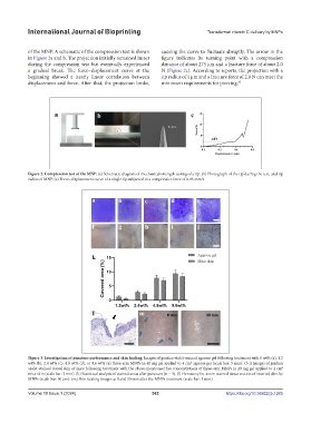
of the MNP. A schematic of the compression test is shown causing the curve to fluctuate abruptly. The arrow in the
in Figure 2a and b. The projection initially remained intact figure indicates its turning point with a compression
during the compression test but eventually experienced distance of about 275 μm and a fracture force of about 2.0
a gradual break. The force–displacement curve at the N (Figure 2c). According to reports, the projection with a
beginning showed a nearly linear correlation between tip radius of 4 μm and a fracture force of 2.0 N can meet the
displacement and force. After that, the projection broke, minimum requirements for piercing. 32
Figure 2. Compression test of the MNP. (a) Schematic diagram of mechanical strength testing of a tip. (b) Photograph of the tip during the test, and tip
radius of MNP. (c) Force–displacement curve of a single tip subjected to a compressive force of 0.05 mm/s.
Figure 3. Investigation of puncture performance and skin healing. Images of gentian violet-stained agarose gel following treatment with 0 wt% (a), 1.2
wt% (b), 2.4 wt% (c), 4.8 wt% (d), or 9.6 wt% (e) three-arm MNPs in 40 mg gel applied to 4 cm agarose gel (scale bar: 5 mm). (f–j) Images of gentian
2
violet-stained dorsal skin of mice following treatment with the above-mentioned five concentrations of three-arm MNPs in 20 mg gel applied to 2 cm 2
mice skin (scale bar: 2 mm). (k) Statistical analysis of stained areas after puncture (n = 3). (l) Hematoxylin–eosin-stained tissue section of inserted skin by
MNPs (scale bar: 50 μm). (m) Skin healing images at 0 and 30 min after the MNPs treatment (scale bar: 5 mm).
Volume 10 Issue 1 (2024) 362 https://doi.org/10.36922/ijb.1285