
Page 469 - IJB-10-1
P. 469
International Journal of Bioprinting TPMS bone scaffold
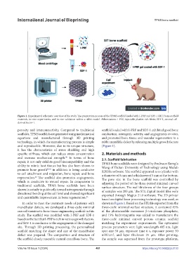
Figure 1. Experimental schematic overview of the study. The preparation process of the TPMS scaffold loaded with I-PRF and SDF-1 (SIT) bone scaffold
materials, in vitro experiments, and in vivo validation within a rabbit model. Abbreviations: I-PRF, injectable platelet-rich fibrin; SDF-1, stromal cell-
derived factor-1.
porosity and interconnectivity. Compared to traditional scaffold loaded with I-PRF and SDF-1 exhibited good bone
scaffolds, TPMS scaffolds are generated using mathematical conduction, osteogenic activity, and angiogenesis in vitro,
equations and manufactured through 3D printing and promoted bone tissue and vascular regeneration in a
technology, in which the manufacturing process is simple rabbit mandible defect by releasing multiple growth factors
and reproducible. Moreover, due to its unique structure, (Figure 1).
it has the characteristics of stress shielding and high
specific stiffness, which can reduce stress concentration 2. Materials and methods
40
and increase mechanical strength. In terms of bone 2.1. Scaffold fabrication
repair, it not only exhibits good biocompatibility and the TPMS bone scaffolds were designed by Professor Shengfa
ability to mimic host tissues but has also been shown to Wang of Dalian University of Technology using Matlab
promote bone growth 40,41 in addition to being conducive R2020a software. The scaffold appeared as a cylinder with
to cell attachment and migration, bone repair, and bone a diameter of 8 mm and a thickness of 5 mm at the bottom.
42
regeneration. The scaffold also promotes angiogenesis, The pore size in the bone scaffold was controlled by
which is conducive to wound repair. In comparison to adjusting the period of the three-period minimal curved
traditional scaffolds, TPMS bone scaffolds have been surface structure. The wall thickness of the four groups
shown in a study to guide cells toward osteogenesis through of samples was 200 µm. The STL digital model files were
directional bending at the cell level and showed significant exported through Magics 21.0 software. The 3D printer
and quantifiable improvement in bone regeneration. 43
based on digital laser processing technology was used, as
In order to meet the treatment needs of patients with shown in Figure 1. Based on the STL file exported from the
mandibular defects, we developed a three-cycle minimal three-cycle minimal surface structure, it contained 85%
curved biomimetic bone tissue engineering scaffold in this of the photocurable monomer β-tricalcium phosphate,
study. The scaffold was modified with I-PRF and SDF-1 and 15% hydroxyapatite was added to manufacture the
based on the fact that I-PRF is rich in various growth factors, three-cycle minimal curved porous ceramic scaffold
and SDF-1 is conducive to the angiogenesis of the damaged matching the experiment requirements. The optimized
site. Through 3D printing processing, the personalized process parameters were light wavelength 405 nm, light
scaffold matching the shape and size of the mandibular spot size 50 µm, exposure time 6 s, exposure power 30
defect was prepared. The composition and structure of mW/cm , and layer thickness 25 µm. After printing,
2
the scaffold closely resemble natural cancellous bone. The the sample was separated from the prototype platform,
Volume 10 Issue 1 (2024) 461 https://doi.org/10.36922/ijb.0153