
Page 63 - IJB-2-1
P. 63
Wei Long Ng, Wai Yee Yeong and May Win Naing
Figure. 6. (Left) Excessive material deposition, (Middle) optimal printing parameters, (Right) incomplete printing (scale bar: 500 µm).
while a low printing pressure would result in incom- plates followed by seeding of HFF-1 cells onto the
plete patterning. Among all the PGC hydrogels and surface and a control set-up with 2.5% chitosan was
different combinations of printing pressures and feed used in this study. The seeded HFF-1 cells were gen-
rates, the 5% PGC hydrogels at printing conditions of erally round in shape and evenly distributed over the
2.4 bars and 1000 mm/min feed rate enables the fa- surface of PGC hydrogels. Live-dead staining was
brication of complete grid-like patterns at highest conducted to visually inspect the cell viability and
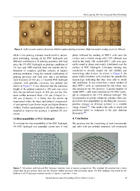
printing resolution. Using the optimal combination of morphology after 4 days. As shown in Figure 8, the
printing pressures and feed rates and a pre-defined green viable fibroblast cells exhibited the spindle-like
layer thickness of 160 μm, a 3-layered PGC hydrogel morphology indicating that they were able to attach
construct with grid-like structures was printed and and proliferate. It was noted that a small number of
shown in Figure 7. It was observed that the estimated dead HFF-1 cells, as represented by red colour, was
height of the printed construct (~400 μm) was lower also present in the 3D construct. A greater number of
than the pre-defined height of 480 μm and the fila- viable HFF-1 cells were observed on 5% PGC hydro-
ment widths increased from ~314 μm (1-layer) to ~ gel as compared to the 2.5% chitosan hydrogel. The
450 μm (3-layers). It is likely that the nozzle tip incorporation of gelatin within the polymer blend im-
transversed within the layer and induced compression proved the biocompatibility by shielding the excessive
of each printed layer (lower height and higher filament positive charges on chitosan polymer to a suitable
widths). Further optimization to the layer thickness is charge density [30] . This enables the cells to attach and
required to improve the accuracy of printed 3D con- proliferate better as compared to the pure chitosan
structs. biomaterial [40] .
3.4 Biocompatibility of PGC Hydrogels 4. Conclusion
To evaluate the biocompatibility of the PGC hydrogel, We envision that the bioprinting of both biomaterials
5% PGC hydrogel was manually casted onto 6 well and cells with pre-defined structures will eventually
Figure 7. 3D printed multi-layered PGC hydrogel structure view at various persepectives. The resultant 3D construct has a lower
height than the pre-defined value and the filament widths increased with increasing layers. 5% PGC hydrogel was tested for the
fabrication of 3D hydrogel construct, number of layers 3, scale bar = 5 mm
International Journal of Bioprinting (2016)–Volume 2, Issue 1 59