
Page 10 - IJB-2-2
P. 10
Advancing cancer research using bioprinting for tumor-on-a-chip platforms
money (US $2.6 billion) [34,35] , there is a need for al- tissue architectures with ease [39–42] . The technology
ternative options in preclinical drug testing [36] . A low- offers high throughput and excellent reproducibility,
cost, reproducible model that mimics tumors, includ- generating cancer tissue models which closely mimic
ing the microenvironment, cell distribution and vas- the structure and function of tumors in vivo, including
culature, would allow high-throughput drug screening tumor heterogeneity and vascular structures. With
prior to clinical trials as an efficient alternative to an- rapid advances in bioprinting technology for cancer
imal models. Such a bioprinted model has already models, there is potential to expand our basic under-
[5]
been reported for cervical cancer . Additionally, bio- standing of cancer and develop effective therapies.
printed models can be used to test other materials re-
levant to drug delivery, such as scaffolds for releasing Conflict of Interest and Funding
signals [37] and polymer microspheres for biodegrada-
tion studies [38] . No conflict of interest was reported by the authors. ST
Although there is room for further innovation in acknowledges the University of Connecticut Research
bioprinting, this approach shows great promise for Excellence Program award for financial support of
efficient generation of biomimetic tumor models to this research.
further advance and accelerate cancer research. A Acknowledgements
unique advantage of bioprinting compared to other
microfabrication techniques is the ability to precisely The authors would like to acknowledge Chu Hsiang
control the spatial arrangement of cells and complex Yu for preparing the figure in this article.
(A) (C)
(B)
(D)
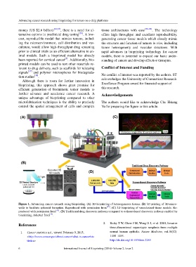
Figure 1. Advancing cancer research using bioprinting. (A) 3D bioprinting of heterogeneous tissues. (B) 3D printing of 3D micro-
wells to facilitate spheroid formation. Reproduced with permission from [25] . (C) 3D bioprinting of vascularized tissue models. Re-
produced with permission from [43] . (D) Traditional drug discovery pathway compared to a tissue-based discovery pathway enabled by
bioprinting. Adapted from [44] .
References 2. Ridky T W, Chow J M, Wong D J, et al. 2010, Invasive
three-dimensional organotypic neoplasia from multiple
1. Cancer statistics n.d., viewed February 9, 2015, normal human epithelia. Nature Medicine, vol.16(12):
<http://www.cancer.gov/about-cancer/what-is-cancer/sta 1450–1455.
tistics> http://dx.doi.org/10.1038/nm.2265
6 International Journal of Bioprinting (2016)–Volume 2, Issue 2