
Page 15 - IJB-2-2
P. 15
Dhakshinamoorthy Sundaramurthi, Sakandar Rauf and Charlotte A. E. Hauser
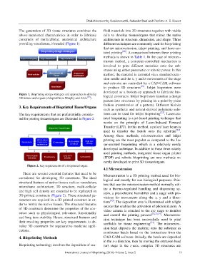
The generation of 3D tissue structures combines the ffold materials into 3D structures together with viable
above mentioned characteristics in order to fabricate cells to develop tissues/organs that mimic the native
constructs of multicellular, anatomical architecture architecture in structure, dimension, and shape. Three
providing vasculature, if needed (Figure 1). different techniques are commonly used for bioprinting
that are microextrusion, inkjet printing, and laser-ass-
isted printing [20] . A comparison between these printing
methods is shown in Table 1. In the case of microex-
trusion method, a computer-controlled mechanism is
involved to print different materials onto the sub-
strates using either pneumatic or robotic power. In this
method, the material is extruded via a standard extru-
sion needle and the x, y and z-movements of the stage
and extruder are controlled by a CAD-CAM software
to produce 3D structures [21] . Inkjet bioprinters were
developed as a bottom-up approach to fabricate bio-
Figure 1. Bioprinting design strategies and approaches to develop
3D tissues and organs (Adopted from Murphy and Atala [19] ). logical constructs. Inkjet bioprinters translate a design
pattern into structures by printing in a point-by-point
3. Key Requirements of Bioprinted Tissue/Organs fashion (rasterization of a pattern). Different bioinks
such as synthetic and natural-derived polymeric solu-
The key requirements that are preferentially conside- tions can be used for inkjet bioprinting [22] . Laser-ass-
red for printing tissues/organs are illustrated in Figure 2. isted bioprinting is a jet-based printing technique that
works on the principle of Laser-Induced Forward
Transfer (LIFT). In this method, a pulsed laser beam is
used to transfer the bioink onto the substrate [23] .
Among these methods, microextrusion and inkjet
printing are the most popular as compared to the La-
ser-assisted bioprinting which is a relatively newly
developed technique. In addition to these three widely
used printing methods, integrated tissue organ printer
(ITOP) and robotic bioprinting are new methods re-
cently developed to print 3D tissues/organs.
Figure 2. Key requirements of a bioprinted organ.
4.1 Microextrusion
There are several essential features that need to be Microextrusion is a 3D printing method used for bio-
considered for developing 3D constructs. The ideal logical and mostly for non-biological purposes. Prin-
structural features of native tissues such as vasculature, ters that use the microextrusion method normally util-
micro/nano architecture, 3D structure, multi-cellular ize a thermo-regulated handling and dispensing sy-
and high cell density are essential to be replicated in stem, a piezoelectric humidifier and a stage with pro-
3D printed constructs (Figure 2). These structural pa- visions for movements along the x, y and z direc-
rameters are required in a 3D printed construct in or- tions [33] . The deposition area is illuminated with a light
der to mimic the native tissues. The structural features source that enables the activation of photoinitiators. A
of 3D constructs determine the properties of the con- video camera is attached to the xyz stage to monitor
struct such as physiological relevance, functionality and control the printing process [18,33,34] . Microextru-
and long term stability. Hence, structural features and sion technique has been successfully used to print
their resulting properties are key requirements to de- scaffolds for tissue engineering [34] . The microextru-
velop 3D constructs for regenerative medicine appli- sion head deposits the material onto the substrate as
cations.
continuous beads based on the instructions from the
4. Bioprinting Methods CAD-CAM software. Initially, the beads are deposited
in the x-y direction, then by moving the extrusion head
Bioprinting technology involves the deposition of sca- (or) stage in the z-axis, complex 3D structures are
International Journal of Bioprinting (2016)–Volume 2, Issue 2 11