
Page 18 - IJB-2-2
P. 18
3D bioprinting technology for regenerative medicine applications
this type of printers require bioink of fast gelation ki- tion and oxygen. Recently, ITOP (Integrated Tissue
netics to develop constructs with good shape fidelity, Organ Printer) bioprinting method has been reported
and this may hinder the flow rate during printing [25,58] . for the fabrication of complex human tissues with
Also, preparing multiple cells containing ribbon or good viability and vasculature [69] . This approach dem-
absorbing layer is a time-consuming process. The con- onstrated the printing of various polymers and cell ty-
structs that are fabricated by laser-assisted bioprinting pes in a single tissue construct using multi-dispensing
are often found to contain traces of contamination modules. ITOP uses pneumatic-actuated microextru-
(comes from the absorbing layer). To avoid such con- sion method but differ in dispensing systems, hard-
taminations absorbing layers made up of non-metallic ware and software as discussed below. ITOP method
substances are being used [59] . Furthermore, it is hard uses air pressure to control dispensing volume and a
to focus the laser spot and to precisely locate the cells three-axis motorized stage for 3D patterning. The 3D
during printing. To overcome this difficulty, the “aim patterns employed in ITOP method were generated
and shoot” technique is used [60] . Here, the laser beam from computed tomography (CT) and magnetic re-
will scan and choose the region of interest, in order to sonance imaging (MRI) data of human organs/tissues.
locate specific cells and to eject one cell per laser This data was finally converted into 3D patterns using
pulse [60] . Using this printing method bone constructs a computer-aided design (CAD) software. It was pro-
and skin with cells for implantation have been suc- posed that ITOP method can offer many advantages
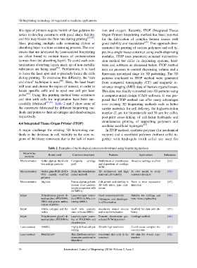
cessfully fabricated [61,62] . Table 2 and 3 show some of over existing 3D bioprinting methods such as better
the constructs fabricated by different bioprinting me- carrier materials for cell delivery, the high-resolution
thods and points to their advantages and disadvantages, nozzles (2 μm for biomaterials and 50 μm for cells),
respectively. post-print cross-linking of cell-laden hydrogels and
simultaneous printing of supporting polymers and
4.4 Integrated Tissue Organ Printer (ITOP)
acellular sacrificial hydrogels [69] .
A major challenge for existing 3D bioprinting me- In ITOP method, synthetic polymer (for mechanical
thods is the decrease in cell viability in the core re- support) and a sacrificial polymer (without cells) to-
gions of the tissue constructs due to the lack of nutri- gether with hydrogels (with cells) are used for
Table 2. Examples of the biological constructs developed using bioprinting methods
Bioprinting Bioink used Construct developed Features Applications References
methods
Microextrusion Gellan-alginate blend with Composite cartilage Proliferation of chondrocytes Bioactive cartilage scaffold [63]
biocartilage particles graft and deposition of cartilage
ECM
Microextrusion Gellan gum-RGD (RGD- Brain-like branched ne- 3D architecture and high In vitro model to study [64]
GG)- peptide modified uronal network neuronal cell viability neuronal disorders
polymer
Microextrusion Alginate Porous alginate gel with Cell growth and viability in Patch to treat myocardial [65]
Human fetal cardiom- 3D with native gene expr- infarction
yocyte progenitor cells ession
(hCMPCs)
Inkjet Poly(ethylene glycol) di- Layer-by-layer assem- Good cytocompatibility. Suitable for cartilage and [66]
methacrylate (PEGDMA, bled PEG-GelMa con- Osteogenic and chodrogen- bone tissue engineering
PEG) and gelatin metha- taining hMSCs ic differentiation
crylate (GelMA)
Inkjet Fibrin, collagen and thr- Graft with amniotic Accelerate wound closure Scaffold for stem cells de- [67]
ombin stem cells and MSCs and re-epithelialization livery
Inkjet Poly(ethylene glycol) di- Layer-by-layer assem- Promote chondrocyte gro- Cartilage scaffold [68]
methacrylate (PEGDMA) bly of PEGDMA and wth and ECM deposition
chondrocytes
Laser-assisted HMSCs Highly defined cell pat- 3D with high resolution Could create complex tis- [61]
terning sue structure
Laser-assisted Matriderm® Skin fibroblasts and ker- Functional skin cells in the 3D skin for wound rege- [62]
atinocytes positioned in construct neration
Matriderm
14 International Journal of Bioprinting (2016)–Volume 2, Issue 2