
Page 16 - IJB-2-2
P. 16
3D bioprinting technology for regenerative medicine applications
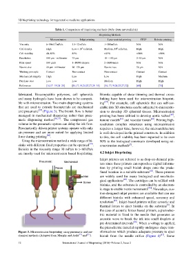
Table 1. Comparison of bioprinting methods (N/A- Data not available)
Bioprinting Methods
Microextrusion Inkjet printing Laser-assisted printing ITOP Robotic printing
7
Viscosity 6–30×10 mPa/s 3.5–12 mPa/s 1–300 mPa/s N/A N/A
6
8
Cell density High Low < 10 cells/mL Medium, 10 cells/mL High High
Cell viability 40–80% 85% >95% >90% >90%
Resolution 100 μm - millimeter 75 μm 10 – 100 μm 2–50 μm N/A
Print speed 100 μm/s 1–10000 drops/s 2–1600 mm/s N/A N/A
Nozzle size 20 μm- millimeter 20–150 μm Nozzle-less 50 μm N/A
Working principle Contact Non-contact Non-contact Contact Contact
Mechanical integrity High Low Low High Medium
Purchase cost Low Low Medium High High
References [14,17–19,24–28] [14,17,19,24,25,27,29–31] [14,17,19,24,25,27,32] [69] [70]
fabricated. Biocompatible polymers, cell spheroids Bioinks capable of shear thinning and thermal cross-
and many hydrogels have been shown to be compati- linking have been used for microextrusion bioprint-
ble with microextrusion. Two main dispensing systems ing [39] . For example, cell spheroids that can self-ass-
that are used to extrude biomaterials are mechanical emble into 3D structures can be subjected to microextru-
and pneumatic [35] (Figure 3). The bioink flow is better sion to develop 3D spheroid tissues. Microextrusion
managed in mechanical dispensing rather than pneu- printing has been utilized to develop aortic valves [40] ,
matic dispensing method [36,37] . The compressed gas tumour models [41] and vascular tissues [42] . Printing high-
volume in the pneumatic system can delay the ink flow. resolution complex structures using microextrusion
Pneumatically driven printer systems operate with only requires a longer time, however, the microarchitecture
air-pressure and are more suited for applying limited is well developed in the printed constructs. In addition
force during printing [20] . to this, the cell viability has been reported to be over
Using the microextrusion method a wide range of bi- 90% in the biological constructs developed using mi-
oinks with different fluid properties can be operated [38] . croextrusion methods .
[5]
Bioinks in the viscosity range 30 mPa/s to > 60 kPa/s
are mostly used for microextrusion based bioprinting. 4.2 Inkjet Bioprinting
Inkjet printers are referred to as drop-on-demand prin-
ters since these printers can reproduce digital informa-
tion by printing small bioink drops onto the prede-
fined location in a suitable substrate [43] . These printers
are widely used for many biological and non-biolo-
gical applications [44] . The cartridges can be refilled with
bioinks, and the substrate is controlled by an electron-
ic stage to enable z-axis movements [45] . Nowadays, cus-
tom-designed inkjet printers are available that can use
different bioinks with enhanced speed, accuracy and
resolution [45] . Inkjet-based printers utilize acoustic and
[5]
thermal forces to eject bioinks on the substrate . In
the case of acoustic forces based printers, a piezoelec-
tric material is fixed to the needle that generates an
acoustic wave to break the ink into small droplets at
pre-determined intervals [46] . When a voltage is applied,
the piezoelectric material rapidly undergoes shape tran-
Figure 3. Microextrusion bioprinting using pneumatic and me- sformations which produce adequate pressure to eject
chanical methods (Adopted from Murphy and Atala [19] and [21] ). bioink from the needle orifice (Figure 4) [47] . Some
12 International Journal of Bioprinting (2016)–Volume 2, Issue 2