
Page 19 - IJB-2-2
P. 19
Dhakshinamoorthy Sundaramurthi, Sakandar Rauf and Charlotte A. E. Hauser
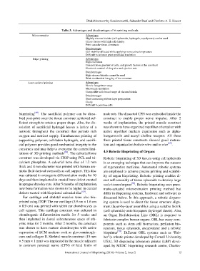
Table 3. Advantages and disadvantages of bioprinting methods
Microextrusion Advantages
Slightly viscous bioinks (cell spheroids, hydrogels, copolymers) can be used
Create tissues with high cell density
Print vascular tissue constructs
Disadvantages
Cell viability affected while applying more extrusion pressure
Difficult to enhance print speed and resolution
Inkjet printing Advantages
High resolution
Concentration gradient of cells, and growth factors in the construct
Electronic control of drop size and ejection rate
Disadvantages
High viscous bioinks cannot be used
Weak mechanical integrity of the construct
Laser-assisted printing Advantages
Nozzle-less printer setup
Microscale resolution
Compatible with broad range of viscous bioinks
Disadvantages
Time consuming ribbon layer preparation
Costly
Difficult to position cells
bioprinting [69] . The sacrificial polymer can be disso- nude rats. The dissected CPN was embedded inside the
lved post-print once the tissue construct achieved suf- construct to enable proper nerve impulse. After 2
ficient strength to retain a proper shape. Also, the dis- weeks of implantation, the printed muscle construct
solution of sacrificial hydrogel leaves a lattice of a was shown to have organized myofiber orientation with
network throughout the construct that permits rich native myoblast markers expression such as alpha-
oxygen and nutrient supply. Simultaneous printing of bungarotoxin and acetyl choline receptor. All these
supporting polymer, cell-laden hydrogels, and sacrifi- three printed tissue constructs showed good matura-
cial polymer provides good mechanical integrity to the tion and organization both in vitro and in vivo [69] .
constructs and may help to overcome the current limi-
tations of 3D printing methods [69] . The calvarial bone 4.5 Robotic Bioprinting of Organs
construct was developed via ITOP using PCL and tri- Robotic bioprinting of 3D tissues using cell spheroids
calcium phosphate. A calvarial bone disc of 1.2 mm is an emerging technique that can improve the success
thick and 8 mm diameter was printed with human am- of regenerative medicine. Automated robotic systems
niotic fluid derived stem cells as cell support. This disc are employed to achieve precise printing and scalabil-
was cultured in osteogenic differentiation media for 10 ity of organ bioprinting. Robotic printing enables di-
days and then implanted in cranial bone defect created rect self-assembly of tissue spheroids to develop large
in sprague drawley rats. After 5 months of implantation, scale tissues/organs [70] . Robotic bioprinting uses pneu-
new bone formation was shown to be higher in cranial matic-actuated microextrusion printing method but
defects treated with bioprinted calvarial disc [69] . differ in dispensing systems, hardware and software as
Ear cartilage and skeletal muscles were also bio- discussed below. In this approach, a robotic dispens-
printed using ITOP. The ear cartilage (3.9 cm × 1.6 cm ing system is used to direct the tissue structure align-
× 0.9 cm) was printed with rabbit ear chondrocytes as ment (layer-by-layer assembly) using a suitable bioink
cell support. This cartilage construct was cultured in (cell spheroids) onto biopapers (hydrogel sheets). Also,
chondrogenic differentiation media for 5 weeks and an Organ Biofabrication Line (OBL) is required to
then implanted in dorsal subcutaneous space of ath- fabricate complex human organs. OBL has many com-
ymic mice for 2 months. After 2 months, the construct ponents such as stem cell bioreactors, perfusion bio-
was shown to have mature chondrocytes with native reactors, tissue spheroids, encapsulator and a robotic
expression of ECM markers such as glycosaminogly- bioprinter [70] . Different OBL systems such as “Fab-
cans and collagen II. Skeletal muscle construct (15 mm ber”(a robotic printer developed by Cornell University,
× 5 mm × 1 mm) was implanted in the muscle adjacent USA), 3D dispensing laboratory printer (LBP) devel-
to common peroneal nerve (CPN) of hind limbs of oped by MUSC bioprinting research centre, Charles-
International Journal of Bioprinting (2016)–Volume 2, Issue 2 15