
Page 23 - IJB-2-2
P. 23
Dhakshinamoorthy Sundaramurthi, Sakandar Rauf and Charlotte A. E. Hauser
meters that need to be considered to develop 3D con- ulus of >500 mPa and used to print bone constru-
structs for regenerative medicine applications. Some cts [104] . In another study, Cooper et al. [105] have devel-
of the applications of 3D bioprinting in regenerative oped a 3D printed bone made from DermaMatix and
medicine are listed in Table 4. BMP-2 (Bone Morphogenetic Protein-2). This con-
Existing skin grafting techniques and commercially struct showed an effective healing of a calvarial defect
available skin grafts do not meet all the requirements in a mouse model [105] .
that are needed for aesthetic skin regeneration. Bio- Bioprinting can be employed to develop neural
printing methods have been employed to construct stem cells constructs to treat central nervous system
complex stratified layers of skin that may be used in (CNS) diseases such as Huntington’s disease, Parkin-
skin grafting applications [62] . Lee et al. [102] have engi- son’s disease, and Alzheimer’s disease. Hsieh et al. [106]
neered skin tissue constructs through a layer-by-layer printed 3D neural tissue constructs of thermorespon-
assembly of collagen, dermal fibroblasts and epider- sive polyurethane containing neural stem cells. These
mal keratinocytes. The printed 3D skin was proposed neural stem cells-laden 3D printed polyurethane scaf-
to be useful as a skin substitute to treat full thickness folds rescued traumatic brain injury in a zebrafish
skin damages [102] . model. Here, bioprinting of neural stem cells was
Bioprinted cartilage may mimic some of the prop- demonstrated to improve neural stem cells encapsula-
erties of the native cartilage and could be useful as a tion and viability.
scaffold for the repair of cartilage damages such as Aortic valves or prosthetic heart substitutes are de-
joint injuries [96,103] . For example, bioink made of PEG veloped for regenerative medicine applications [65,40] .
and alginate can form an interpenetrating network [103] . These biological structures have complex architecture
Additional crosslinking of this hybrid polymer using and also contain multiple cell types. 3D bioprinting
calcium sulfate allowed higher cell encapsulation and methods may offer ways to develop aortic valves/heart
–2
also showed toughness (1500 J m ) greater than na- substitutes with native structure and viable cells. In a
tive cartilage [96,103] . recent study, alginate–gelatin aortic valve was fabri-
Critical-sized bone defects require graft assistance cated using 3D bioprinting [40] . This bioprinted valve
for healing. Although tissue engineered scaffolds offer was claimed to closely match the native anatomy of
solutions to the existing problems associated with aortic valve. In addition to the structural similarity, the
non-healing bone defects, an ideal scaffold that can fabricated aortic valve also has viable aortic smooth
restore the native functions of the bone is yet to be muscle cells and aortic valve leaflet interstitial cells.
identified [104] . Bioprinting methods may provide an These studies demonstrate that it is possible to create
alternative method to the development of bone scaf- aortic valves using 3D bioprinting [40] .
folds that closely mimic the native functions of the Pluripotent stem cells and embryonic stem cells (ES
bone. For example, PEGDMA hydrogel developed via cells) are cell sources for patient-specific treatments
photopolymerization method had a compressive mod- and hence attractive for 3D bioprinting of constructs
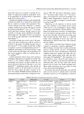
Table 4. Applications of bioprinting in regenerative medicine
Application Bioink Printing method Cell type Inference Ref.
Cartilage Alginate-Polyethylene Microextrusion Bone marrow derived hMSCs Tougher mechanical integrity like [103]
glycol native cartilage
Acrylonitrile butadiene st- Microextrusion Primary articular chondrocy- Porous scaffold for cartilage and [96]
yrene (ABS) and polylac- tes and nucleus pulposus intervertebral disc tissue enginee-
tic acid (PLA) ring
Skin Layer-by-layer assembled Microextrusion Human skin fibroblasts and Skin matrix that resembles struc- [102]
collagen human skin keratinocytes tural and biological features of
native skin
Bone DermaMatrix™ human all- Inkjet Mouse C2C12 progenitor cells Osteogenic differentiation of C2- [105]
ograft with bone morph- C12 cells and promotes clavarial
ogenetic protein-2 bone healing
Nerve Polyurethane Microextrusion Neural stem cells Recovery from CNS neural injury [106]
in zebra fish
Heart Alginate-gelatin Microextrusion Aortic root sinus smooth mus- Cell encapsulated aortic valve re- [40]
cle cells (SMC) and aortic valve tain anatomic complexity
leaflet interstitial cells (VIC)
Liver Alginate Microextrusion Human induced pluripotent Post-print differentiation into hep- [107]
stem cells atocyte lineage
International Journal of Bioprinting (2016)–Volume 2, Issue 2 19