
Page 100 - IJB-10-2
P. 100
International Journal of Bioprinting 3D-printed nanocomposites: Synthesis & applications
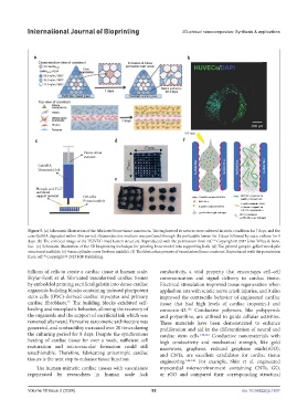
Figure 5. (a) Schematic illustration of the fabricated bone tissue constructs. The engineered structures were cultured in static condition for 7 days, and the
core GelMA degraded within this period. Osteoinductive medium was perfused through the perfusable lumen for 5 days followed by static culture for 9
days. (b) The confocal image of the HUVEC-lined lumen structure. Reproduced with the permission from ref. Copyright © 2017 John Wiley & Sons,
139
Inc. (c) Schematic illustration of the 3D bioprinting technique for printing bone model into supporting bath. (d) The printed genipin-gelled wood-pile
structured scaffolds. (e) 4 mm cylinder cores (bottom middle). (f) The fabrication process of vascularized bone construct. Reproduced with the permission
from ref. Copyright © 2023 IOP Publishing.
140
billions of cells to create a cardiac tissue at human scale. conductivity, a vital property that encourages cell–cell
Skylar-Scott et al. fabricated vascularized cardiac tissues communication and signal delivery in cardiac tissue.
by embedded printing sacrificial gelatin into dense cardiac Electrical stimulation improved tissue regeneration when
organoids building blocks containing induced pluripotent applied on rats with sciatic nerve crush injuries, and it also
stem cells (iPSC)-derived cardiac myocytes and primary improved the contractile behavior of engineered cardiac
cardiac fibroblasts. The building blocks exhibited self- tissue that had high levels of cardiac troponin-I and
45
healing and viscoplastic behavior, allowing the recovery of connexin-43. Conductive polymers, like polypyrrole
142
the organoids and the support of sacrificial ink which was and polyaniline, are utilized to guide cellular activities.
removed afterward. Pervasive sarcomeric architecture was These materials have been demonstrated to enhance
generated, and contractility increased over 20 times during proliferation and aid in the differentiation of neural and
the culturing period for 8 days. Despite the synchronous cardiac stem cells. 142,143 Conductive nanomaterials with
beating of cardiac tissue for over a week, sufficient cell high conductivity and mechanical strength, like gold
maturation and microvascular formation could still nanowires, graphene, reduced graphene oxide(rGO),
unachievable. Therefore, fabricating anisotropic cardiac and CNTs, are excellent candidates for cardiac tissue
tissues is the next step to enhance tissue function. engineering. 144-146 For example, Shin et al. engineered
The human-mimetic cardiac tissues with vasculature myocardial microenvironment containing CNTs, GO,
regenerated by researchers in human scale lack or rGO and compared their corresponding structural
Volume 10 Issue 2 (2024) 92 doi: 10.36922/ijb.1637