
Page 102 - IJB-10-2
P. 102
International Journal of Bioprinting 3D-printed nanocomposites: Synthesis & applications
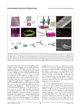
Figure 6. Bioprinting perfused vasculature. (a) Schematic illustration of cardiac patch model and the printing process concept. (b) A continuous layer of
endothelial cells (ECs) inside the printed blood vessels and the cross-sectional view of lumen. Reproduced with the permission from ref. Copyright © 2019
88
John Wiley & Sons, Inc. (c) Schematics of a rotated rod template used in coaxial printing of bioinks (left). A picture of a double-layer vessel-like structure
(top right). Fluorescent image of three cell lines-encapsulated vessels (bottom right). Reproduced with the permission from ref. Copyright © 2017
156
American Chemical Society. (d) The fabrication process of branched micro-channels generated via coaxial printing (left). Scanning electron microscopy
(SEM) image of double-channel part of Y-shaped microchannels after trimming (right). Reproduced with the permission from ref. Copyright © 2016 AIP.
161
physical properties of the vessels, including stiffness, elastic endothelial cells were seeded by perfusion post-printing,
modulus, vascular shrinkage rate, and liquid adsorption HUVECs were mixed with gelatin in this study and printed
capacity. Further, the cell viability was not influenced by in the core of extruded filaments. Endothelial cells deposit
MWCNTs in the short term, but the cell viability, cellular and adhere to inner wall of the microchannels during
motility, and the generation of ECM were reduced in the long static culture period. Gelatin was then dissolved to obtain
term due to CNT-induced toxicity. 158,159 Other researchers vascular channels. Endothelial cells proliferated over
have found that the cell viability in the hollow structure the culturing time and spread and formed a layer on the
was higher than that in constructs without microchannels, inner side of the vascular channels. Moreover, angiogenic
indicating the importance of vessels and nutrients delivery sprouts were observed in the bioprinted constructs, further
in large-scale organ models. Additionally, co-culturing suggesting its in vivo application.
41
HUVECs and MSCs in the bioprinted constructs promoted
cell function of HUVECs, including proliferation, protein The coaxial bioprinting approach can also be used
expression, and angiogenesis. Gelatin is another kind in conjunction with a rotated rod to create hierarchical
43
156
of sacrificial material which can be injected through the architectures as shown in Figure 6c. However, one main
inner nozzle as it can be easily liquefied and removed challenge of this strategy is to fabricate interconnected
157
by increasing temperature. Shao et al. prepared GelMA branched vessels. Li et al. developed a novel method to
bioinks encapsulated with tissue cells that were extruded engineer branched micro-channel via partial crosslinking
161
through the outer nozzle onto a cooling platform allowing and trimming (Figure 6d). The patency of branched
the thermo-crosslinking of gelatin. The printed stacked vessels was confirmed by injecting cell media, and the
160
vessels constructs were then permanently UV-crosslinked. fibroblasts were found to maintain high viability and
Different from the previous published methods that proliferate after being cultured for 6 days. Moreover, vessels
Volume 10 Issue 2 (2024) 94 doi: 10.36922/ijb.1637