
Page 149 - IJB-10-2
P. 149
International Journal of Bioprinting dECM bioink for in vitro disease modeling
time and chemical concentration should be optimized in The main purpose of decellularization is to preserve
an effort to preserve the ECM molecules. In particular, ECM components of the native tissue. The residual ECMs
decellularization methods should be tissue-specific because can be roughly classified as collagens, adhesive proteins,
tissues and organs have different mechanical stiffnesses, and GAGs. The degree of preservation of the ECM is
as well as protein and lipid compositions. A list of tissue- currently a nonstandard yardstick for assessing the utility
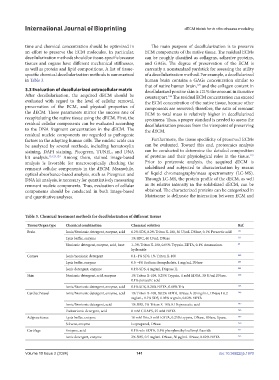
specific chemical decellularization methods is summarized of a decellularization method. For example, a decellularized
in Table 3. human brain contains a GAGs concentration similar to
that of native human brain, and the collagen content in
125
3.3 Evaluation of decellularized extracellular matrix decellularized porcine skin is 121% the amount in its native
After decellularization, the acquired dECM should be counterpart. The residual ECM concentration can exceed
126
evaluated with regard to the level of cellular removal, the ECM concentration of the native tissue, because other
preservation of the ECM, and physical properties of components are removed; therefore, the ratio of remnant
the dECM. These parameters mirror the success rate of ECM to total mass is relatively higher in decellularized
recapitulating the native tissue using the dECM. First, the specimens. Thus, a proper standard is needed to assess the
residual cellular components can be evaluated according decellularization process from the viewpoint of preserving
to the DNA fragment concentration in the dECM. The the dECM.
residual nucleic components are regarded as pathogenic
factors in the adapting human cells. The nucleic acids can Furthermore, the tissue specificity of preserved ECMs
be analyzed by several methods, including hematoxylin can be evaluated. Toward this end, proteomics analysis
staining, DAPI staining, Picogreen, TUNEL, and DNA can be conducted to determine the detailed composition
127
kit analysis. 15,123,124 Among them, stained image-based of proteins and their physiological roles in the tissue.
analysis is favorable for macroscopically checking the Prior to proteomic analysis, the acquired dECM is
remnant cellular components in the dECM. Meanwhile, solubilized and subjected to characterization by means
optical absorbance-based analysis, such as Picogreen and of liquid chromatography/mass spectrometry (LC-MS).
DNA kit analysis, is necessary for quantitatively measuring Through LC-MS, the protein profile of the dECM, as well
remnant nucleic components. Thus, evaluation of cellular as its relative intensity in the solubilized dECM, can be
components should be conducted in both image-based obtained. The characterized proteins can be categorized in
and quantitative analyses. Matrisome to delineate the interaction between ECM and
Table 3. Chemical treatment methods for decellularization of different tissues
Tissue/Organ type Chemical combination Chemical solution Ref.
Brain Ionic/Nonionic detergent, enzyme, acid 0.2% SDS, 0.2% Triton X-100, 50 U/mL DNase, 0.1% Peracetic acid 141
Lysis buffer, enzyme 1% SDC, 40 U/mL DNase 131
Nonionic detergent, enzyme, acid, base 1–3% Triton X-100, 0.05% Trypsin-EDTA, 0.1% Ammonium 85
hydroxide
Cornea Ionic/nonionic detergent 0.1–1% SDS, 1% Triton X-100 242
Lysis buffer, enzyme 0.5 –4% Sodium deoxycholate, 1 mg/mL DNase 243
Ionic detergent, enzyme 0.1% SDS, 4 mg/mL Dispase II, 244
Skin Nonionic detergent, acid, enzyme 1% Triton X-100, 0.25% Trypsin, 1 mM EDTA, 30 U/ml DNase, 126
0.1% peracetic acid
Ionic/Nonionic detergent, enzyme, acid 0.1% SDS, 0.26% EDTA, 0.69% Tris 245
Cardiac/Vessel Ionic/Nonionic detergent, enzyme, acid 1% Triton X-100, 0.02% EDTA, RNase A 20 mg/mL, DNase I 0.2 246
mg/mL, 0.1% SDS, 0.05% trypsin, 0.02% EDTA
Ionic/Nonionic detergent, acid 1% SDS, 1% Triton X-100, 0.1% peracetic acid 247
Zwitterionic detergent, acid 8 mM CHAPS, 25 mM EDTA 248
Adipose tissue Lysis buffer, enzyme 10 mM Tris, 5 mM EDTA, 0.25% trypsin, DNase, RNase, lipase, 249
Solvent, enzyme Isopropanol, DNase 250
Cartilage Enzyme, acid 0.1% w/v EDTA, 3.5% phenylmethyl sulfonyl fluoride 251
Ionic detergent, enzyme 2% SDS, 0.5 mg/mL DNase, 50 μg/mL RNase, 0.02% EDTA 252
Volume 10 Issue 2 (2024) 141 doi: 10.36922/ijb.1970