
Page 192 - IJB-10-2
P. 192
International Journal of Bioprinting Optimizing inkjet bioprinting
as φ. These groups are characterized as follows: dilute ( 12. 5 )
27
bio-inks (with φ ≤ 2%), semi-dilute bio-inks (within 2% ≤ 0 (I)
φ ≤ 25%), and concentrated bio-inks (with φ > 25%). The
typical diameter of mammalian cells is ~16 µm, and when This model is valid for very dilute suspensions of
the cell concentrations are 1, 10, and 120 million cells/ rigid spheres (φ < 0.01), where φ represents the viscosity
mL, this corresponds to volume fractions of 0.21%, 2.15%, of the pure solvent. The model was extended to account
29
and 25.74%, respectively. The introduction of cells into for viscous spheres with internal viscosity η and can be
28
s
30
the suspension leads to an increase in the effective (shear) expressed as:
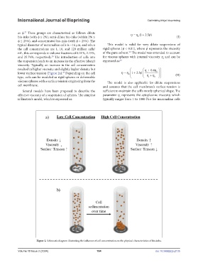
viscosity. Typically, an increase in the cell concentration
resulted in higher viscosity and slightly higher density but 04.
s
5.
lower surface tension (Figure 2a). Depending on the cell 0 12 0 (II)
23
type, cells can be modeled as rigid spheres or deformable s 0
viscous spheres with a surface tension originating from the The model is also applicable for dilute suspensions
cell membrane. and assumes that the cell membrane’s surface tension is
Several models have been proposed to describe the sufficient to maintain the cell’s mostly spherical shape. The
effective viscosity of a suspension of spheres. The simplest parameter η represents the cytoplasmic viscosity, which
s
is Einstein’s model, which is expressed as: typically ranges from 1 to 1000 Pa·s for mammalian cells
Figure 2. Schematic diagram illustrating the influence of cell concentration on the physical characteristics of bio-inks.
Volume 10 Issue 2 (2024) 184 doi: 10.36922/ijb.2135