
Page 196 - IJB-10-2
P. 196
International Journal of Bioprinting Optimizing inkjet bioprinting
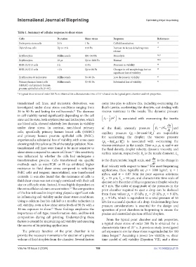
Table 1. Summary of cellular response to shear stress
Cells Duration Shear stress Response References
Embryonic stem cells 24 h 1 Pa Cell differentiation 52
Hybridoma cells Up to 15 h 0.16 Pa Increase in lactate dehydrogenase 53
release
Erythrocytes Milliseconds 450–560 Pa Hemolysis 56,57
Erythrocytes 10 µs Up to 4000 Pa Normal 58,59
BHK-21/C13 cells 2 h 10 Pa Decrease in viability 60
BHK-21/C13 cells <1 h Up to 80 Pa Changes in cell morphology, but no 60
significant loss of viability
Erythrocytes & leukocytes Milliseconds 30–90 Pa Low decrease in viability 61
Primary human breast cells Milliseconds 30–90 Pa Substantial loss of viability 61
(HMEC) and primary human
prostate epithelial cells (PrEC)
*A typical shear stress of order 300 Pa is obtained for a characteristic time of 10 s based on the typical print chamber and ink properties.
-5
transformed cell lines, and metastatic derivatives, was come into play to achieve this, including overcoming the
investigated under shear stress conditions ranging from fluid’s inertia, accelerating the droplets, and dealing with
30 to 90 Pa and lasting for milliseconds. The decrease viscous resistance in the nozzle. The dynamic pressure
61
in cell viability varied significantly depending on the cell 1 2
state and its state; both erythrocytes and leukocytes, which p u is associated with overcoming the inertia
d
2
are blood cells, showed relatively low decrease in viability du
under shear stress. In contrast, non-blood primary of the fluid; unsteady pressure p L n and
a
dt
cells, specifically primary human breast cells (HMEC) capillary pressure (p 2 cos / R ) are responsible
c
n
and primary human prostate epithelial cells (PrEC), for accelerating the droplet; the viscous pressure
experienced a substantial loss of viability, with some cases (p 8η L uR 2 ) is associated with overcoming the
/
n
n
v
showing viability as low as 2% of the total population. Non- viscous resistance in the nozzle. Here a ρ, µ, η, and σ are
transformed cell lines were found to be more sensitive to the fluid density, droplet velocity, dynamic viscosity, and
shear stress compared to cancer cell lines. This sensitivity surface tension, respectively; R is the nozzle diameter, L
61
was influenced by whether the cells had undergone a n n
transformation process. Cells transformed via specific is the characteristic length scale, and du is the change in
dt
methods such as myc/PI3K or H-ras exhibited higher fluid velocity with respect to time. For most bioprinting
62
resistance to fluid shear stress compared to wild-type applications, these typically are ρ = 1000 kg/m , η = 1
3
PrEC cells and isogenic immortalized, non-transformed mPa·s, and σ = 0.07 N/m for pure aqueous solutions;
controls. It was also found that the resistance of cells to R = 15 µm, L = 50 µm, and characteristic time scale of
n
n
fluid shear stress was not strongly correlated with their cell ejection is in the order of 10 µs to generate a droplet velocity
size or cell cycle state. Instead, it was highly dependent on of 5 m/s. The order of magnitude of the pressures in the
the extracellular calcium concentration. The composition print chamber required to eject a drop can be deduced
61
of the bio-ink used in bioprinting played a significant role from these values: p = 13 kPa, p = 25 kPa, p = 9 kPa,
c
d
a
in influencing cell viability under shear stress conditions. p = 9 kPa, which is equivalent to a total pressure of 56
v
Using a calcium-free bio-ink led to a notable reduction in kPa for successful ejection of a drop. Understanding these
cell viability, even at low shear stress levels of 50 Pa with a pressure considerations is essential for the design and
10 ms exposure to shear. These findings emphasize the operation of print chambers in bioprinting to ensure the
61
importance of cell type, transformation state, and bio-ink precise and controlled ejection of fluid droplets.
composition during cell printing. Understanding these
From the typical print chamber and ink properties,
factors is crucial for maintaining cell viability and ensuring a typical shear stress of order 300 Pa is obtained for a
the success of bioprinting applications.
characteristic time of 10 s. A previous study investigated
-5
The primary function of the print chamber is to cell exposure to similar shear stress magnitudes but for 100
provide the necessary momentum for ejection of a precise times longer. Assuming a power law viability vs. shear
61
volume of fluid droplets from the chamber. Several factors time model of cell viability (Equation VII), and a shear
Volume 10 Issue 2 (2024) 188 doi: 10.36922/ijb.2135