
Page 28 - IJB-3-1
P. 28
Digital biomanufacturing supporting vascularization in 3D bioprinting
of neovessels, whether by angiogenesis or vasculoge- 5.3 Biomanufacture of Vascularized Systems
nesis. This is true of those materials in which vascular
cells are incorporated at the time of fabrication (i.e., a With the promise of these in vitro microcirculations,
hydrogel) or rigid scaffolds that are made and subse- exciting new opportunities arise for building more
quently seeded with vascular cells or vascular ele- native-like tissue models and mimics for use in the
ments [32,33] . Moreover, many of the native matrices laboratory (and eventually tissue replacement). How-
used as bioinks have intrinsic pro-angiogenic activity ever, these vascularization advances raise new bio-
such as tumor matrix and hyaluronic acid gels [34,35] . Of manufacturing challenges as the complexity of the
course, many strategies have doped bioinks with an- systems rise. For example, individual cell types within
giogenic factors either to drive vasculogenesis/angio- systems such as endothelial cells, other vascular cells,
genesis from embedded vascular precursors and/or targeted parenchymal cells (e.g., hepatocytes, tumor
recruit vessel ingrowth into the construct via angioge- cells), and tissue-specific stromal cells all have unique
nesis. The different materials used promote vascular media and microenvironmental requirements that
adaptation to different degrees with softer, native ma- must be coordinated to support the construct as a
trices being the most favored. Rigid scaffolds do sup- whole. Also, new biofabrication strategies addressing
port vascular adaptation, however, this relies on the when and how to integrate vasculatures with paren-
spaces between the rigid elements, where the neoves- chyma cells and other cells types, including staged
sels reside, being filled with a softer material. incubation steps, need to be developed. While 3D bi-
oprinting is a key fabrication approach, the successful
5.1 Synthetic Channels strategies in the future will undoubtedly include other
fabrication methods. Related to this, organizing man-
An alternate approach to incorporating a perfusion
supply involves creating channels through a matrix ufacturing workflows becomes paramount as different
within which vascular cells (usually endothelial cells) fabrication steps are staged through the entire manu-
facturing process. While these practices are common
are seeded onto the channel walls, thereby fabricating to non-biological manufacturing programs, their ap-
a simple vessel-like element. Connecting the channels plications to biomanufacturing have yet to be com-
to each other results in a perfusable network of endo- prehensively implemented (Figure 1). However, new
thelial cell-lined channels serving to provide a means tools enabling these broader biomanufacturing activi-
fluid flow through the construct. The endothelial cell ties with living systems are emerging [36] and groups
lining adds a biological dynamic to the channels by are beginning to develop the concepts and methods
functionalizing the fluid-tissue interface as a regulated needed to build complex tissues.
exchange barrier. However, adaptation into more na-
tive-like microvasculatures is limited as the channel
topology is fixed and vascular remodeling, even with
the addition of other vascular cells is constrained.
Cellularized channel systems are usually made ei-
ther by soft photolithographic methods or 3D bio-
printed sacrificial reverse molds [36] .
5.2 Combining Approaches
The latest efforts at establishing a microcirculation in
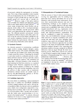
vitro seeks to combine the microfluidic endothelial Figure 1. Example work flow (arrows) for digital biomanufac-
cell-lined channel platform with a native, derived mi- turing. A medical scan of a patient (an MRI of the chest) is
crovasculature. Here, the channels serve as a perfusion imported directly into a commercially available prototyping
software (TSIM®, Advanced Solutions Life Science). The bio-
source which, when contiguously connected to a neig- logical content (i.e., the structure to be fabricated) is extracted
hboring microvasculature, help to drive the formation from the image set to produce a 3D digital prototype which is
of a microcirculation. Often, vascular cells are used to then used to print the physical version in a contour-printing
form the initial, native microvasculature to be con- capable robotic arm printer (BioAssembly Bot®, Advanced
nected to the channel system [37] . In contrast, Advanced Solutions Life Science). The process entails a spectrum of
technologies including image processing, in silico model gen-
Solutions Life Sciences is using isolated microvessels eration, biology, clinical data, 3D prototyping, robotics, bioma-
to form the native microcirculation [36] . terials, and cell biology.
24 International Journal of Bioprinting (2017)–Volume 3, Issue 1