
Page 55 - IJB-3-1
P. 55
Lothar Koch, Ole Brandt, Andrea Deiwick, et al.
(A) (B)
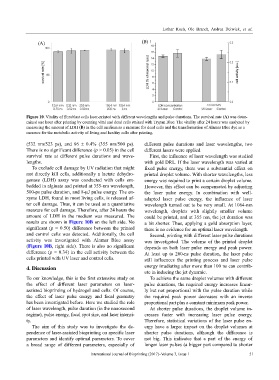
Figure 10. Vitality of fibroblast cells laser-printed with different wavelengths and pulse durations. The survival rate (A) was deter-
mined one hour after printing by counting vital and dead cells stained with Trypan Blue. The vitality after 24 hours was analyzed by
measuring the amount of LDH (B) in the cell medium as a measure for dead cells and the transformation of Alamar Blue dye as a
measure for the metabolic activity of living and healthy cells after printing.
(532 nm/523 ps), and 96 ± 0.4% (355 nm/500 ps). different pulse durations and laser wavelengths, two
There is no significant difference (p > 0.05) in the cell different lasers were applied.
survival rate at different pulse durations and wave- First, the influence of laser wavelength was studied
lengths. with gold DRL. If the laser wavelength was varied at
To exclude cell damage by UV radiation that might fixed pulse energy, there was a substantial effect on
not directly kill cells, additionally a lactate dehydro- printed droplet volume. With shorter wavelengths, less
genase (LDH) assay was conducted with cells em- energy was required to print a certain droplet volume.
bedded in alginate and printed at 355-nm wavelength, However, this effect can be compensated by adjusting
500-ps pulse duration, and 8-µJ pulse energy. The en- the laser pulse energy. In combination with well-
zyme LDH, found in most living cells, is released af- adapted laser pulse energy, the influence of laser
ter cell damage. Thus, it can be used as a quantitative wavelength turned out to be very small. At 1064-nm
measure for cell damage. Therefore, after 24 hours the wavelength, droplets with slightly smaller volume
amount of LDH in the medium was measured. The could be printed, and at 355 nm, the jet duration was
results are shown in Figure 10B on the left side. No a bit shorter. Thus, applying a gold absorption layer,
significant (p = 0.50) difference between the printed there is no evidence for an optimal laser wavelength.
and control cells was detected. Additionally, the cell Second, printing with different laser pulse durations
activity was investigated with Alamar Blue assay was investigated. The volume of the printed droplet
(Figure 10B, right side). There is also no significant depends on both laser pulse energy and peak power.
difference (p = 0.34) in the cell activity between the At least up to 200-ns pulse duration, the laser pulse
cells printed with UV laser and control cells. still influences the printing process and laser pulse
energy irradiating after more than 100 ns can contrib-
4. Discussion
ute in inducing the jet dynamic.
To our knowledge, this is the first extensive study on To achieve the same droplet volumes with different
the effect of different laser parameters on laser- pulse durations, the required energy increases linear-
assisted bioprinting of hydrogel and cells. Of course, ly but not proportional with the pulse duration while
the effect of laser pulse energy and focal geometry the required peak power decreases with an inverse
has been investigated before. Here we studied the role proportional part plus a constant minimum peak power.
of laser wavelength, pulse duration (in the nanosecond At shorter pulse durations, the droplet volume in-
regime), pulse energy, focal spot size, and laser intensi- creases faster with increasing laser pulse energy.
ty. Therefore, statistical variations of the laser pulse en-
The aim of this study was to investigate the de- ergy have a larger impact on the droplet volumes at
pendence of laser-assisted bioprinting on specific laser shorter pulse durations, although the difference is
parameters and identify optimal parameters. To cover not big. This indicates that a part of the energy of
a broad range of different parameters, especially of longer laser pulses (a bigger part compared to shorter
International Journal of Bioprinting (2017)–Volume 3, Issue 1 51