
Page 50 - IJB-3-1
P. 50
Laser-assisted bioprinting at different wavelengths and pulse durations with a metal dynamic release layer: A parametric study
(EOS 450D, Canon, Krefeld, Germany), stroboscopic was added to the cells. After 4 hours of incubation, the
illumination with a flashlamp (Nanolite KL-M, absorbance of the solution was measured at 570 nm,
High-Speed Photo-System, Wedel, Germany) with 11 with a reference wavelength of 600 nm on a micro-
nanoseconds flash duration, and microscope objective plate reader.
(M Plan Apo NIR 20x, Mitutoyo, Neuss, Germany). A
full description of this setup has been published be- 3. Results
fore [15] .
3.1 Wavelength Variation
2.9 Determination of the Survival Rate
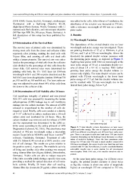
The dependence of the printed droplet size on laser
The survival rate of printed cells was determined by wavelength and pulse energy was investigated. There
rinsing most cells from the donor and collector slides are printing thresholds of 12 µJ at 1064-nm, 6 µJ at
separately after printing, staining the dead cells with 532-nm, and 3 µJ at 355-nm wavelengths. Above the
Trypan Blue, and counting all cells and dead cells threshold, the printed droplet volume increases with
within a hemocytometer. The survival rate was calcu- the increasing pulse energy, as depicted in Figure 3.
lated as the percentage of vital cells from the collector Applying laser pulses with 1064 nm wavelength at the
slide divided by the percentage of vital cells from the laser pulse energy of 30 µJ, a maximum droplet vol-
donor slide. Cell survival rates were determined for ume of about 2.4 ± 0.4 nL is reached. With further
printing with both, the Yb: YAG laser (at 1064-nm growing laser pulse energy the droplet volume in-
wavelength with 8- and 200-ns pulse duration) and the creases only slightly. The same droplet volume can be
Nd:YAG laser (wavelength/pulse duration: 1064 nm/750 printed with 532-nm wavelength at the lower laser
ps; 532 nm/523 ps; 355 nm/500 ps). The laser pulse en- pulse energy of 17.5 µJ, but this droplet volume can-
ergy was adjusted to print about 50% of the cells from not be reached with 355-nm wavelength due to the
the donor to the collector slide. limited laser pulse energy, below 8 µJ.
2.10 Determination of Cell Viability after 24 hours
Cell membrane integrity of printed and non-printed
NIH-3T3 cells was assessed by measuring the lactate
dehydrogenase (LDH) leakage due to cell membrane
damage into the culture medium. The amount of LDH
released is proportional to the number of cells da-
maged or lysed. Briefly, cells were seeded at a density
4
of 5×10 cells/well in culture medium into a 24-well
culture plate and incubated for 24 hours. Then, the
culture medium was removed and the release of LDH
into the supernatant was determined by the LDH ac-
tivity assay according to the online protocol of OPS Figure 3. Printed droplet volume dependence on laser wave-
Diagnostics (Lebanon, NJ, USA). The absorbance was length and pulse energy. The size of alginate droplets printed
detected at 492-nm wavelength using a microplate with three different wavelengths and different laser pulse ener-
TM
reader (Tecan Infinite M200Pro and Tecan i-control gies. There is an upper droplet size limit (near 350-µm droplet
software, Crailsheim, Germany). Treatment of cells diameter) depending on the hydrogel layer thickness as a limi-
with 1% Triton-X100 served as a 100% positive con- tation of available hydrogel. Since the laser maximum pulse
energy for 355 nm (and 532 nm) is much smaller compared to
trol of cell damage. The results are given relative to 1064-nm wavelength, the upper droplet size limit has not been
the positive control, in percent. The metabolic activity reached with 355 nm. Nevertheless, it can be concluded that the
of living and healthy cells after printing was assayed achievable droplet size is quite similar for all investigated
using Alamar Blue dye (Sigma-Aldrich, Deisenhofen, wavelengths. With 1064 nm, a bit smaller droplets can be
printed.
Germany). Viable cells are able to reduce resazurin
(blue) into resorufin (pink) during a specific time span, At the same laser pulse energy, but different wave-
providing a method for optical detection of cell meta- lengths, the printed droplet diameter is about twice
bolic activity. Briefly, 20 hours after cell seeding, as big (the volume is about one order of magnitude
Alamar Blue dye (resazurin 20 µg/mL culture medium) higher) at wavelength 355 nm compared to 532 nm,
46 International Journal of Bioprinting (2017)–Volume 3, Issue 1