
Page 22 - IJB-3-2
P. 22
Bioprinting of osteochondral tissues: A perspective on current gaps and future trends
native osteochondral tissues individually as well as the nicity, may be more of a challenge to treat. Smaller acute
[9]
2
interface region . The heterogeneous and anisotropic lesions (less than 2 cm ) are filled with type-I collagen
cartilage is generally considered as a layered structure fibrocartilage, which has been proven biomechanically
[15]
of “zones” that possess mechanical properties reflecting and histologically inferior to native hyaline cartilage .
2
each zone’s compositional and architectural make- Larger lesions (greater than 2 cm ) require addressing the
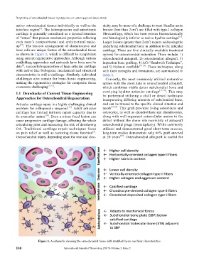
up [10] . The layered arrangement of chondrocytes and underlying subchondral bone in addition to the articular
bone cells are unique feature of the osteochondral tissue cartilage. There are five clinically available treatment
as shown in Figure 1, which is difficult to recapi tulate options for osteochondral restoration. These include: 1)
using current regenerative approaches. Although various osteochondral autograft, 2) osteochondral allograft, 3)
scaffolding approaches and materials have been used to impaction bone grafting, 4) ACI “Sandwich Technique”,
[5]
date , successful regeneration of large articular cartilage and 5) biphasic scaf folds [16–26] . These techniques, along
with native-like biological, mechanical and structural with their strengths and limitations, are summarized in
characteristics is still a challenge. Similarly, individual Table 1.
challenges also remain for bone-tissue engineering, Currently, the most commonly utilized restorative
making the regenerative strategies for composite tissue option with the most data is osteochondral allograft,
even more challenging [11,12] . which combines viable donor subchondral bone and
1.1 Drawbacks of Current Tissue Engineering overlying hyaline articular cartilage [17,18] . This may
Approaches for Osteochondral Rege neration be performed utilizing a shell or dowel technique
incorporating differing amounts of subchondral bone,
Articular cartilage repair is a highly challenging clinical and can be titrated to the specific clinical situation and
problem for orthopaedic surgeons [13] . Adult articular needs [18,24] . This graft provides living osteoblasts and
cartilage has limited intrinsic repair capacity due to osteocytes, as well as chondroblasts and chondrocytes,
its avascular nature [14] . Even a minor focal lesion can along with well-organized extracellular matrix to the
cause progressive cartilage damage, affecting the whole defect without the donor site morbidity of autograft
articulating joint and increasing the risk of developing osteochondral plugs (mosaicplasty). While commonly
OA. Traditional cartilage repair techniques focus utilized and demonstrated good short-term success,
on pain relief as well as restoring tissue function . long-term studies demonstrate only 66% graft survival
[5]
Osteochondral injury, depending upon the size and chro- at 20 years [27] . Osteochondral allograft is useful for
Figure 1. A schematic showing the osteochondral tissue with stratified layers and their characteristics
110 International Journal of Bioprinting (2017)–Volume 3, Issue 2