
Page 23 - IJB-3-2
P. 23
Pallab Datta, et. al.
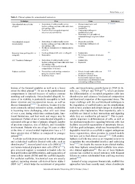
Table 1. Clinical options for osteochondral restoration
Technique Pros Cons References
Osteochondral osteoarticular • Restoration of architecturally correct • Disease transmission and [16–18,24,27,95]
allograft hyaline cartilage and bone with viable bone immunogenicity
and cartilage cells • Availability
• Excellent short-term patient reported • Contouring challenges in
outcomes and survivability patellofemoral joint
• Short-term clinical results not
sustained through long-term follow-
up
Osteochondral autograft • Restoration of architecturally correct • Donor site morbidity [15–18]
(mosaicplasty) hyaline cartilage and bone with viable bone • Contouring challenges in all
and cartilage cells locations
• No disease transmission or immunogenicity
concerns
Impaction bone grafting (allo- or • Can be performed with auto- or allograft • Limited data [23]
autograft) • Inexpensive • No biologic restoration of articular
surface
ACI “Sandwich Technique” • Restoration of architecturally correct • Limited data [96,97]
hyaline cartilage and bone with viable bone • Expensive
and cartilage cells
• Contouring can be modified to all
compartments of the knee
Biphasic scaffolds • Directed bone and cartilage restoration • Mixed clinical results with even [25,26]
using bio-directive matrix short-term follow-up
• Relatively cheap • Breakdown products from
bioresorbable materials may be
chondrotoxic and detrimental to the
surrounding cartilage and bone.
lesions of the femoral condyles as well as to a lesser cells, and transforming growth factor β (TGF-β) fa-
[24]
extent the tibial plateau . Its use in the patellofemoral mily (i.e., TGF-β1 and TGF-β3 [34] ), which promotes
compartment is limited because of surface contour the differentiation of recruited progenitor cells into
matching and complexity. Osteochondral allograft, be- chondrocytes and enhances biochemical composition
cause of its viability, is particularly susceptible to both and functional properties of the regenerated tissue. The
donor rejection and incorporation issues, as well as major challenge with the scaffold-based techniques is
disease transmission [16,18,24] . In addition, because it is the the degradation of scaffold matrix and its complications
most commonly utilized restorative option, availability such as toxic products and abrupt changes in mechanical
is becoming more challenging, often with wait times properties after implantation. Most importantly, cells in
of over a year, during which time continued pain, func- scaffolds are limited in their interactions and signaling
tional limitations, and lost work and wages may be while they are confined in gel matrix [35] . This is parti-
experienced. Failure of use of osteochondral allografts is cularly important in differentiation of cells, as well as
associated with age at time of primary allograft, number the mechanotransductive signaling between cells that
of previous surgeries, size of defects, and bicondylar facilitate successful regeneration of anisotropic tissues.
in volvement. Patients who are of age 30 years or older Cell-free graft techniques, which use biocompatible and
at the time of osteochondral implantation have a 3.5 degradable materials as a scaffold to support endogenous
times greater risk of failure as compared to younger tissue regeneration, show promise in animal models
patients [24,27] . but have yet to find clinical success [36–38] . Cell-seeded,
A cellular scaffolds (more practical in clinical settings) biphasic scaffolds may serve as an integrated solution to
and cellularized scaffolds (loaded with cells, such as reca pitulate the osteochondral interface and underlying
chondrocytes [28] , mesenchymal stem cells (MSCs) [29,30] bone [37,39] , but despite the success in pre-clinical stu dies,
and human-induced pluripotent stem cells (iPSCs) [31,32] ), only three biphasic osteochondral scaffolds have exten-
give structural support to recruited or loaded cells for sive clinical application [40] . The use of these biphasic
their proliferation and differentiation, and regeneration of systems has resulted in mixed outcomes with frequent
extra-cellular matrix (ECM) until the scaffold degrades. failures to restore subchondral bone and long recovery
For acellular scaffolds, biochemical cues are usually periods [40–42] .
applied, including stromal cell-derived factor alpha 1 Instead of using exogenous biomaterials, scaffold-free
(SDF-1α) , which helps the recruitment of progenitor approaches have been recently used, where chondrocyte
[33]
International Journal of Bioprinting (2017)–Volume 3, Issue 2 111