
Page 25 - IJB-3-2
P. 25
Pallab Datta, et. al.
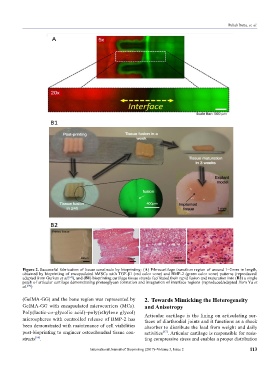
Figure 2. Successful fabrication of tissue constructs by bioprinting: (A) Fibrocartilage transition region of around 1–2 mm in length,
obtained by bioprinting of encapsulated hMSCs with TGF-β1 (red color zone) and BMP-2 (green color zone) patterns (reproduced/
adapted from Gurkan et al. ), and (B1) bioprinting cartilage tissue strands facilitated their rapid fusion and maturation into (B2) a single
[64]
patch of articular cartilage demonstrating proteoglycan formation and integration of interface regions (reproduced/adapted from Yu et
al. )
[94]
(GelMA-GG) and the bone region was represented by 2. Towards Mimicking the Heterogeneity
GelMA-GG with encapsulated microcarriers (MCs). and Anisotropy
Poly(lactic-co-glycolic acid)–poly(ethylene glycol) Articular cartilage is the lining on articulating sur-
mi cro spheres with controlled release of BMP-2 has faces of diarthrodial joints and it functions as a shock
been demonstrated with maintenance of cell viabilities absorber to distribute the load from weight and daily
post-bioprinting to engineer osteochondral tissue con- activities . Articular cartilage is responsible for resis-
[67]
structs . ting compressive stress and enables a proper distribution
[66]
International Journal of Bioprinting (2017)–Volume 3, Issue 2 113