
Page 6 - IJB-3-2
P. 6
Post-printing surface modification and functionalization of 3D-printed biomedical device
substrate is often unheeded, despite the fact that they transformed post-printing. At macroscopic level,
are in constant interactions with biological entities. shape memory materials are incorporated in the
Even for 3D-printed tissue engineering scaffold which 3D-printed components [23–25] . Under the designed
is designed to promote cell attachment and growth, transition conditions (e.g., temperature or solvent), the
its biological functions largely rely on the intrinsic shape memory materials deform and transform the
property of the bulk materials, which imposes fur- 3D-printed components into desired configuration.
ther constraints on the already limited selection of 3D- This concept, which is also known as 4D print-
printable materials. ing [26,27] , creates a dynamic component that allows
For 3D-printed components to achieve full potential users to reconfigure the shape of 3D-printed compo-
in biomedical applications, a multiprocess 3D print- nents on demand. We consider the reconfiguration a
ing [22] that combines 3D printing and post-3D printing post-3D printing modification method. The 4D print-
modification is highly coveted. The 3D-printed com- ing technique has been demonstrated for soft robot-
ponents could benefit substantially from the post-3D ics [28] . But we could expect more sophisticated bio-
printing modification for improved biofunctionality. In medical applications using 4D printing technology.
this paper, we first summarize current approaches For example, 4D printing is well-suited for custo-
used for post-3D printing modification. Meanwhile, mized vascular stent. Once the pre-configured stent
we identify the limitations of 3D-printed components reaches the stricture, it could be induced to expand and
for biomedical applications, and provide a perspective open the stenotic vessel to restore blood flow.
on how to close the gap using post-3D printing modi- The post-3D printing architectural reconfigura-
fication techniques. tion could also enhance the performance of 3D-printed
microfluidic devices. Microfluidic devices printed
2. Post-3D Printing Modification
using conventional 3D-printing techniques such as
Post-printing modification has two major effects. It is stereolithography (SLA) or fused deposition modeling
able to reconfigure the 3D architecture and/or chemi- (FDM) have fairly poor lateral resolution. A more ad-
cally functionalize the surface of 3D-printed compo- vanced technique, such as two-photon polymerization
[29]
nents, as shown in Figure 1. (2PP), is able to achieve sub-micro resolution , but
it suffers from low throughput. So far, there is no
3D-printing technology that fabricates microfluidic
devices with both high throughput and high resolution.
Post-3D printing reconfiguration provides a potential
solution. Earlier work by Khine et al. demonstrated a
method to achieve high resolution lithography by cre-
ating large patterns and subsequently shrinking the
device to reduce the pattern size (Figure 2) [30] . The
same concept can be adopted in 3D printing. By print-
ing a 3D microfluidic device with pre-stressed materi-
al and subsequently subjecting it to controlled shrink-
ing, the reconfigured device could achieve high reso-
lution despite the fact that the original device may
have large features with a poor resolution.
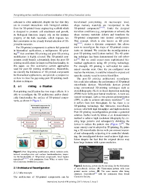
Figure 1. Post 3D-printing modification, which includes archi-
tectural reconfiguration and surface functionalization, impro-
ves biofunctionality of 3D-printed components. Inset figures
reproduced [9,15,17] with permission from Wiley or under Crea-
tive Commons Attribution License.
Figure 2. Reducing pattern size by shrinking for the fabrication
2.1 Architectural Reconfiguration of microfluidic devices. (A) Unshrunken pattern with laser
printed master pattern. (B) The same master after being
2.1.1 Macroscopic shrunken. Reproduced [30] with the permission from Royal
The architecture of 3D-printed components can be Chemistry Society.
94 International Journal of Bioprinting (2017)–Volume 3, Issue 2