
Page 211 - IJB-10-3
P. 211
International Journal of Bioprinting Sr on GO enhances PLLA/PGA scaffold
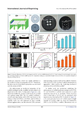
Figure 3. Schematic illustration of SLS (a), the designed model (b), and the scaffold fabricated by SLS (c). Tensile testing (d), tensile strength–strain curves
(e), and tensile strengths of all tested scaffold groups (f). Compression testing (g), compressive strength–strain curves (h), and compressive strengths of
all tested scaffold groups (i).
mechanical properties, which was mainly attributed to with increasing content of GPSr in the scaffolds. However,
the excellent mechanical properties of GO. Meanwhile, GPSr observed on the LG/GPSr2 scaffold showed obvious
62
the improvement of mechanical properties was associated aggregation, which justifies the weakening mechanical
with the content of GPSr. properties of the LG/GPSr2 scaffold. 63
The enhancement in mechanical properties of the To further study the mechanism underlying the
scaffold resulting from the nanofiller was also related to its enhancement of scaffold mechanical properties by GPSr,
dispersion in the matrix. As depicted in Figure 4a–e, the LG and LG/GPSr1.5 scaffolds were subjected to crack
dispersion of different contents of GPSr in the LG scaffold expansion induced by an automatic Vickers hardness
was explored using SEM. The LG scaffold exhibited a tester at the load of 9.8 N, and the generated crack
smooth and clean surface, while GPSr was found on the expansion was observed by SEM. As shown in Figure 4f–
surfaces of the LG/GPSr0.5, LG/GPSr1, and LG/GPSr1.5 i, the crack generated in the LG scaffold passed straight
scaffolds. GPSr was well dispersed on these scaffolds through the scaffold, while the crack was hindered by
without agglomeration, and the observed GPSr increased GPSr in the LG/GPSr1.5 scaffold. Phenomena such as
Volume 10 Issue 3 (2024) 203 doi: 10.36922/ijb.1829