
Page 227 - IJB-10-3
P. 227
International Journal of Bioprinting Preparation and characterization of branched NGCs
Figure 1c, all the DBNs and MBNs were unobstructed the in vivo effects of different branch angles on branched
and could be filled with the dye solution. Meanwhile, the nerve regeneration (Figure 2a). The rat sciatic nerve
degradable behaviors of the conduits were also evaluated defect serves as a primary model for peripheral nerve
in vitro. The results indicated that the degradation rate of regeneration research. This model is chosen due to
the hybrid hydrogel was 10% in 144 h in a 1% collagenase I its appropriate length, diameter, elongated direction,
solution (Figure S4 in Supplementary File). In our previous and adequate space for implanting bulk nerve grafts,
study, we demonstrated that GelMA/PEGDA conduits making it an ideal fit for our study. However, it has been
exhibit biodegradability. In this study, with a higher reported that using a silicone Y-shaped chamber may
51
concentration of PEGDA, the GelMA/PEGDA hydrogel was result in selective regeneration of motor and sensory
biodegradable, although the degradation rate was slower. nerves. 52,53 When a branched conduit is implanted to
bridge a branched nerve gap, the bifurcating pathways
3.2. Nerve regeneration in dual-branched NGCs connect to different distal nerve stumps, potentially
DBNs with predetermined angles were surgically resulting in varied outcomes in nerve regeneration
implanted into the sciatic nerve gap of rats to investigate due to differences in the microenvironment between
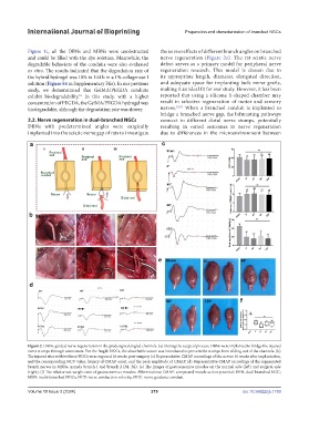
Figure 2. DBNs-guided nerve regeneration in the predesigned angled channels. (a) During the surgical process, DBNs were implanted to bridge the injured
nerve stumps through connectors. For the fragile NGCs, the absorbable suture was introduced to prevent the stumps from sliding out of the channels. (b)
The injured sites with/without NGCs were exposed 16 weeks post-surgery. (c) Representative CMAP recordings of the nerves 16 weeks after implantation,
and the corresponding NCV value, latency of CMAP onset, and the peak amplitude of CMAP. (d) Representative CMAP recordings of the regenerated
branch nerves in MBNs, namely branch 1 and branch 2 (B1, B2). (e) The images of gastrocnemius muscles on the normal side (left) and surgical side
(right). (f) The relative wet weight ratio of gastrocnemius muscles. Abbreviations: CMAP: compound muscle action potential; DBN: dual-branched NGC;
MBN: multi-branched NGCs; NCV: nerve conduction velocity; NGC: nerve guidance conduit.
Volume 10 Issue 3 (2024) 219 doi: 10.36922/ijb.1750