
Page 228 - IJB-10-3
P. 228
International Journal of Bioprinting Preparation and characterization of branched NGCs
the two pathways. 54,55 To eliminate the influence of on the branched nerves (B1 and B2) (Figure 2d).
different nerve stumps, a linear sciatic nerve defect Unexpectedly, there were no significant differences in
model was established in this study, creating a single electrophysiological function among the trunk and
microenvironment induced by a single injured distal branches, specifically B1 and B2. The result hinted that
stump for nerve regeneration. the branched nerves within dual-branched channels
At the 16-week postoperative mark, the functional exhibit comparable electrophysiological conduction.
recovery of the implanted sites and NGCs was evaluated. Our findings suggested that branch angles did not
The distal and proximal stumps of the injured nerves significantly impact nerve regeneration within DBN
were successfully reconnected in all groups (Figure 2b), when the branched conduits were implanted to bridge
and nerve regeneration along the branched channels with linear sciatic nerve gaps. Furthermore, we assessed the
various angles was observed in all DBNs. In the DBNs gastrocnemius muscle’s relative wet weight ratios (i.e., the
with a branch angle of 120°, a segment of the regenerated weight of the right side divided by that of the left side).
nerves even displayed an elongating direction that The results showed no significant differences among
was reversed to the distal stumps to a certain degree DBN groups (Figure 2e–f). Histological analysis of
(Figure S5 in Supplementary File). Electrophysiology gastrocnemius muscle cross-sections was also performed.
examinations were performed to assess the physiological The mean diameters of muscle fibers were similar among
functions of the regenerated nerves. As shown in Figure all the branched NGC groups (Figure 3g; Figure S7 in
2c, no significant difference was observed in NCV and Supplementary File). These results were consistent with
CMAP latency among all DBN groups. However, the electrophysiology assessments.
peak amplitude of CMAP in the sham group exhibited The regenerated branched nerves, bifurcating from the
a significantly lower value than that of the DBN groups. proximal stump and converging toward the distal stumps,
The recovery of CMAP amplitude indirectly reflects were observed (Figure 3a). Histological examination
the number of regenerated motor nerve fibers, while through H&E staining confirmed the regenerated nerves,
a reduced level of CMAP amplitude may suggest the showcasing the presence of newly formed capillaries
incomplete innervation of target tissues. 56,57 Besides, (Figure 3b). Concurrently, LFB staining of myelin
the electrophysiological assessment was also performed sheaths served to further validate the remyelination
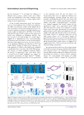
Figure 3. Histological analysis of nerves in DBNs. (a) Representative photographs of the fixed nerves with DBNs (scale bar = 5 mm). (b) H&E staining
(scale bar = 500 μm) and (c) LFB staining of the regenerated nerve tissues in conduits (scale bar = 200 μm). (d) H&E staining and LFB staining of the
longitudinal section of the regenerated branched nerves in DBNs (120°) (scale bar = 1 mm). (e) H&E staining of the regenerated branched nerves (B1,
B2) (scale bar = 200 μm). (f) The relative cross-section area ratio of regenerated branched nerves. (g) The diameter of muscle fibers. Abbreviations: DBN:
dual-branched NGC; H&E: hematoxylin and eosin; LFB: luxol fast blue.
Volume 10 Issue 3 (2024) 220 doi: 10.36922/ijb.1750