
Page 229 - IJB-10-3
P. 229
International Journal of Bioprinting Preparation and characterization of branched NGCs
of the regenerated axons (Figure 3c; Figure S6 in at acute, right, and even obtuse angles to establish
Supplementary File). Notably, myelination was observed connections with the distal end.
in the DBN groups and the sham group. The histological
analysis of nerve longitudinal sections in DBN (120°) 3.3. Nerve regeneration in multi-branched NGCs
further indicated the formation of myelinated nerve To investigate the effect of branch number on branched
branches (Figure 3d). In addition, the assessment of the nerve regeneration, we utilized MBN implantation to
relative cross-section area ratio, calculated in terms of the bridge a linear defect of rat sciatic nerve (Figure 4a).
cross-section area of nerve B1 to that of nerve B2, yielded Following a 16-week period, the two injured nerve stumps
results indicating no significant differences among all were reconnected. Electrophysiology analysis (Figure
the branched NGC groups (Figure 3e–f). In summary, 4c and d) revealed no significant differences in the NCV
the results demonstrated that peripheral nerves exhibit and the latency of CMAP between the MBN group and
the capability to elongate into dual branches in DBNs. the single-channel NGC group (0°). Meanwhile, the wet
Furthermore, these nerves exhibited growth patterns weight ratio of the gastrocnemius muscle demonstrated a
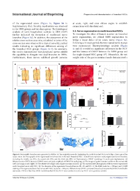
Figure 4. Nerve multi-branches were generated in MBNs. (a) The surgical process for implanting the multi-branched NGCs to bridge a linear gap of the
sciatic nerve. (b) Photographs of the regenerated nerve branches in the multi-branched NGC. Dotted boxes represent newly regenerated nerve branches,
including the middle one (B1), the second side (B2), and the third side (B3). (c) Representative CMAP recordings 16 weeks post-implantation. (d)
Measurements of NCV, latency of CMAP onset, and peak amplitude of CMAP for each group. (e) The images of gastrocnemius muscles on the normal side
(left) and surgical side (right). (f) The relative wet weight ratio of gastrocnemius muscles. (g) The representative photographs of the harvested regenerated
multi-branched nerve with NGC. (h) The diameter of the regenerated branched nerves (B1, B2, and B3). (i) The H&E (scale bar = 500 μm) and (j) LFB
staining of the regenerated branched nerves with NGC (scale bar = 500 μm). Abbreviations: CMAP: compound muscle action potential; H&E: hematoxylin
and eosin; LFB: luxol fast blue; MBN: multi-branched NGCs; NCV: nerve conduction velocity; NGC: nerve guidance conduit.
Volume 10 Issue 3 (2024) 221 doi: 10.36922/ijb.1750