
Page 421 - IJB-10-3
P. 421
International Journal of Bioprinting Expanding 3D cell proliferation with DLP bioprinting
derived GelMA, considerations of religious restrictions shift of the δ = 1.8 portion of the blue (c) functional group
or limitations due to mammalian infectious diseases, is due to the decrease in the methyl group (3H) (Figure 2B).
and a lower melting point, which enables large-scale Successful methacrylation of gelatin resulted in a decrease
synthesis. In addition to GelMA, a variety of other in the chemical shift of the red (b) functional group at δ =
41
photocurable polymers (e.g., methacrylated collagen, 2.9 ppm, which is assigned to the lysine methylene (2H)
methacrylated alginate, methacrylated hyaluronic acid, protons (Figure 2B). To calculate the DoF of F-gelatin,
methacryloyl-modified silk, and methacrylated chitosan) the decrease in the lysine spectrum resulting from the
have been utilized and studied for 3D printing using formation of a new functional group due to the reaction
photopolymerization in recent years. 12,42 between gelatin and MA was targeted. Based on these
54
Achieving high-resolution and biocompatible 3D results, the DoF of F-GelMA was confirmed to be 95.6 ±
bioprinting requires selecting appropriate photoinitiator 2.8% (Figure 2B).
concentration and wavelength. The photoinitiator LAP, 3.1.2. Photorheological analysis of bioink
with its fast speed and high-resolution printability, has Rheological analysis of the bioink was performed to
been utilized in DLP printing as a chemical covalent determine the optimal exposure time for successful DLP
crosslinker of polymers with biocompatible properties and bioprinting. Gelation time, which is the minimum exposure
absorption at ultraviolet (UV) and blue light wavelengths. time, can affect the cellular microenvironment and cell
43
Recently, the range of wavelengths used in DLP printing has viability. Therefore, determining the appropriate exposure
31
expanded to include long wavelengths, such as green light time and applying it to printing enable sophisticated, high-
(500–580 nm) and near-infrared wavelengths (900–1000 resolution printing. The bioink was measured using a
nm), to improve tissue penetration, cell viability rates, rheometer with a gap distance of 100 μm, and the phase
and printing resolution. 44,45 Furthermore, the addition of a transition was explored by examining the storage modulus
light-absorbing agent during DLP printing can help resolve (G’) and loss modulus (G’’). Before photopolymerization,
any mismatches between the bioink polymer and the light G’ was lower than G’’, indicating solution-state dominance,
wavelength, thereby improving printing accuracy. Previous which lasted for up to 27 s. Once photopolymerization
studies in our laboratory compared eight colors of light- occurred under 405 nm visible light, a rapid increase was
absorbing agents used in DLP printing and found that observed in both G’ and G’’, which resulted in a crossover
yellow (405 nm) had the most suitable peak at a UV-Vis
wavelength of 380–450 nm. Tartrazine, a yellow pigment, point indicating the gelation time. This point represents
31
was added to the bioink to prevent excessive crosslinking the occurrence of the solution-to-gel phase change, and
and improve printability. The cells encapsulated in the the gelation time was determined to be 31.13 s. However,
bioink were bovine ear-derived fibroblasts. Fibroblasts, between 27 and 29 s, there was a sharp decrease in G’, which
a type of cell that synthesizes ECM and collagen, are may have been caused by the coexistence of solution and gel
widely recognized as the primary cellular component of and the inaccurate measurement values by the rheometer
31,55
connective tissue in animals. 46,47 They also play pivotal due to uneven polymer formation. After the completion
roles in the formation of the structural skeleton and wound of photopolymerization, G’’ was significantly higher than
healing in animals. 48-50 Therefore, they offer the advantage G’, and there was a tendency for both to rapidly increase
of mimicking the dermis layer of the skin and have the due to the continuous crosslinking reaction. The printable
potential for application as cell-cultivated meat and cultured window highlighted in yellow shows the range of optimized
56
leather. In addition, our laboratory has been conducting printing times after gelation (Figure 2C). The optimal
6,37
basic research on cell-cultivated meat and the utilization of exposure time required for bioink photocrosslinking using
GelMA scaffolds; therefore, we utilized bovine ear-derived 3D DLP printing was determined to be 31.13 s.
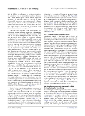
fibroblasts for the current research 6,51 (Figure 2A). 3.2. Morphological analysis of printed F-GelMA
The DoF of the F-gelatin molecules was determined by hydrogel using DLP bioprinting
analyzing the H NMR spectra. 52,53 F-Gelatin and F-GelMA For analyzing the morphological characteristics of printed
1
contain a wide range of amino acids, and their complex F-GelMA 3D hydrogel cross-sections, bioink was printed
spectra can be used to determine the DoF of the molecules. at 100 μm intervals (Figure 2D-(a)). The resulting samples
F-GelMA showed the emergence of a distinct yellow (a) were prepared by cutting the sections after freeze-drying
functional group, while the red (b) and blue (c) functional (Figure 2D-(b)). The porous structure of the resulting
groups decreased compared to F-gelatin (Figure 2A). The hydrogel scaffold was observed and analyzed via SEM
chemical shift of the δ = 5.4/5.7 ppm portion of the yellow (Figure 2D-(c)). The resulting structure using the DLP
(a) functional group is attributed to the acryl protons (2H) stacking method had a layered structure with 100 μm
of the methacrylate group and increases, while the chemical spacing, and multiple pore structures were observed
Volume 10 Issue 3 (2024) 413 doi: 10.36922/ijb.2219