
Page 418 - IJB-10-3
P. 418
International Journal of Bioprinting Expanding 3D cell proliferation with DLP bioprinting
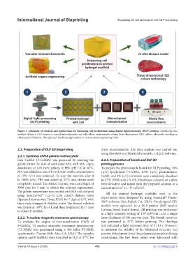
Figure 1. Schematic of research and applications for enhancing cell proliferation using digital light processing (DLP) printing. Specifically, this
method utilizes a DLP printer to control microchannels and cell culture environments during three-dimensional (3D) culture, ultimately resulting in
enhanced proliferation. This approach has broad applications in various tissue engineering fields.
2.2. Preparation of DLP 3D bioprinting these measurements. The data analysis was carried out
using MestReNova (Mestrelab research, v14.2.1) software.
2.2.1. Synthesis of fish gelatin methacrylate
Fish GelMA (F-GelMA) was prepared by reacting the 2.2.3. Preparation of bioink and DLP 3D
gelatin (from the skin of cold-water fish) with MA. Upon printing process
dissolution of 10% (w/v) gelatin in PBS (pH 7.5) at 50°C, To prepare the photocurable bioink for DLP printing, 10%
MA was added at a rate of 0.5 mL/min until a concentration (w/v) lyophilized F-GelMA, 0.5% (w/v) photoinitiator
of 15% (v/v) was achieved. To stop the reaction after 4 (LAP), and 1% (v/v) tartrazine were completely dissolved
h, 100% (v/v) PBS was added at 45°C and stirred until in 37°C DPBS with 1% P/S. Fibroblasts cultured on a plate
completely mixed. The stirred mixture was centrifuged at were detached and mixed with the prepared solution at a
3000 rpm for 5 min to obtain the solution supernatant. concentration of 2 × 10 cells/mL.
6
The gelatin supernatant was reacted with MA and dialyzed All the printed hydrogel scaffolds used in the
using Spectra/Por (12–14 kDa cutoff) dialysis tubes experiments were designed by using Autodesk Fusion
®
®
(Spectra Laboratories, Texas, USA) for 7 days at 45°C with ®
three daily changes of distilled water. The filtered solution 360 software (San Rafael, CA, USA). Pre-designed STL
was frozen at -80°C for 4 h and then lyophilized for 7 days models were uploaded to a DLP printer (IM2 model;
to obtain F-GelMA. Carima, Seoul, South Korea). All printing was performed
at a light intensity setting of 1.97 mW/cm , and a single
2
2.2.2. H nuclear magnetic resonance spectroscopy layer thickness of 100 µm was used. The bioink reservoir
1
To evaluate the degree of functionalization (DoF) of was preheated to 37°C before printing. The rheology
F-GelMA, H nuclear magnetic resonance spectroscopy test indicated a light exposure time of 31.13 s. However,
1
( H-NMR) was performed using a 500 MHz FT-NMR to maintain the stability of the fabricated structure and
1
spectrometer (Varian, Palo Alto, CA, USA). The samples prevent detachment from the polymerization plate during
(gelatin and F-GelMA) were dissolved in D O at 37°C for crosslinking, the first three layers were fabricated with
2
Volume 10 Issue 3 (2024) 410 doi: 10.36922/ijb.2219