
Page 478 - IJB-10-3
P. 478
International Journal of Bioprinting Stretchable scaffold for modeling fibrosis
as a function of patient sex, age, and pathophysiological 2. Methods
conditions of the left ventricle. 22,23 In contrast, diastolic 2.1. Materials
elastic deformation ranges at 10–22%. 21–23 The designed PCL pellets with a molecular weight of 43,000 Da
mechanically stretchable bioartificial scaffolds supported were purchased from Polysciences GmbH (Germany).
the in vitro 3D culture of HCFs. Dynamic in vitro cell GelMA (60% degree of methacryloyl substitution and
culture studies using MechanoCulture T6 bioreactor a gel strength of 300 g Bloom), lithium phenyl-2,4,6-
displayed markers of cardiac fibrosis that could suggest trimethylbenzoylphosphinate (LAP), and 3,4-dihydroxy-
HCFs activation into myofibroblasts. This work highlighted DL-phenylalanine (DOPA) were purchased from Sigma-
the potential of stretchable bioartificial scaffolds for human Aldrich (USA). Phosphate buffered saline (PBS) was
cardiac fibrotic tissue engineering by in vitro culture of purchased from Thermo Fisher Scientific (USA), while
HCFs under cyclic mechanical stimulation. In comparison HCFs and fibroblast growth medium-3 (FGM-3) were
to miniaturized cardiac fibrotic tissue models on a chip, purchased from PromoCell GmbH (Germany).
29
which are limited to the in vitro preclinical testing of 2.2. Poly(ε-caprolactone) mesh geometry
drugs and nanotherapeutics, the present cardiac scar A computer-aided design (CAD) model of the scaffold was
model could also be exploited for the testing of medical designed based on eight superimposed layers (0.15 mm
devices and advanced therapies based on scaffolds/ thickness per layer) (Figure 1). The geometry was based
hydrogels under dynamic mechanical stimulation. on the alternation of two superimposed layers: the first
Future applications by our group will include the in vitro layer made of wavy equidistant filaments aligned along
preclinical study of cardiac regenerative therapies under the x-direction and the other layer consisting of straight
dynamic conditions, such as the direct reprogramming of parallel filaments aligned along the y-direction (Figure 1).
HCFs into cardiomyocytes. 24,30,31 The wavy pattern was composed of a sequence of half
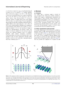
Figure 1. Poly(ε-caprolactone) (PCL) mesh geometry. (A) The geometry of a stretchable PCL mesh unit cell model. Red arrows in the x-direction indicate
the direction of mechanical stretching applied to the mesh; the element portion considered for structural analysis is highlighted in red; spacing dimensions,
expressed in mm, and radius (R) are reported. (B) Single filament cross-section and schematic representation of the superimposition of layers 1 and 2.
(C) The 2D mesh geometry with layers 1 and 2. (D) The 3D mesh geometry is obtained by the superimposition of the eight layers (from layers 1 to 8),
corresponding to four of the 2D mesh structures in (C).
Volume 10 Issue 3 (2024) 470 doi: 10.36922/ijb.2247