
Page 486 - IJB-10-3
P. 486
International Journal of Bioprinting Stretchable scaffold for modeling fibrosis
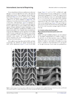
Force values below yield stress conditions were obtained images (Figure 7C and D) of PCL scaffolds with eight
through a numerical algorithm for each of the imposed layers demonstrated that the scaffold geometry accurately
displacement points. Linear regression of the data was reproduced the designed CAD model. The images revealed
performed (Figure 6A), and the stiffness of PCL scaffolds an inter-fiber distance of close wavy and straight filaments
with different thicknesses was computed as the angular of 1.15 ± 0.12 and 1.63 ± 0.07 mm, respectively, compared
coefficient of the curves displayed in Figure S2A. The to the corresponding theoretical values of 1.5 and 2 mm,
evaluation of Von Mises stresses also enabled the validation respectively, in the CAD model. ImageJ analysis of the
of the elastic field hypothesis. Figure 6C illustrates an cross-section optical images (Figure 7B) reported that the
exemplary representation of Von Mises stress distributions cross-section of joint wavy filaments had an average width
for PCL scaffolds with two layers. Scaffolds with a different (b) of 0.22 ± 0.02 mm and an average height (h) of 0.43 ±
number of layers exhibited a similar distribution of Von 0.02 mm.
Mises stresses. Qualitative considerations could be derived
from the stress field of the 1D model: stress was distributed 3.2. Gelatin methacryloyl hydrogels:
along the wavy filaments and the imposition of a bonded Physicochemical characterization and in vitro
contact between straight and wavy filaments concentrated cell tests
stress in proximity of such constraints regardless of the GelMA hydrogel crosslinking kinetics was first analyzed
extinction zones present. The straight filaments were by rheological characterization of GelMA solutions in
mostly unstressed and acted as load propagators in the the presence of LAP photoinitiators using two different
direction of the deformation. light sources (UV and Vis). The sol-gel transition of the
PCL scaffolds were then fabricated by MEX, and their GelMA_5, GelMA_7, and GelMA_10 samples (occurring
morphology was analyzed by optical microscopy and when G’ exceeds G’’) was measured at 37°C during UV
SEM. Optical microscopy (Figure 7A and B) and SEM or Vis light irradiation. Strain sweep tests performed on
Figure 7. Optical images of the (A) top surface and (B) cross-section of an eight-layered PCL scaffold. SEM images of the (C) top surface and (D) tilted
view of a three-layered PCL scaffold. Abbreviations: PCL: Poly(ε-caprolactone); SEM: Scanning electron microscopy.
Volume 10 Issue 3 (2024) 478 doi: 10.36922/ijb.2247