
Page 488 - IJB-10-3
P. 488
International Journal of Bioprinting Stretchable scaffold for modeling fibrosis
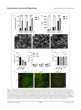
Figure 8. Gelatin methacryloyl (GelMA) hydrogel characterization: (A) crosslinking time and (B) plateau onset values of storage modulus (G’) for GelMA_5,
GelMA_7, and GelMA_10 hydrogels, exposed to ultraviolet (UV) and visible (Vis) light, measured from time-sweep curves (1% strain amplitude and 1 Hz
frequency). ****p < 0.0001. (C–E) Scanning electron microscopy (SEM) images of freeze-dried samples: (C) GelMA_5, (D) GelMA_7, and (E) GelMA_10. (F)
Pore size distribution of the freeze-dried samples and (G) weight loss percentage of GelMA_5, GelMA_7, and GelMA_10 hydrogels incubated in PBS at 37 °C.
(H) Viability (CellTiter-Blue®) of cultured human cardiac fibroblasts (HCFs) in a culture medium containing eluates from GelMA_5, GelMA_7, and GelMA_10
hydrogels after 24 h incubation. (I) Live/Dead Cell Viability assay (green: live cells, red: dead cells) performed on HCF-cellularized GelMA hydrogels cultured
for 24 h and cured accordingly (i.e., 30 s for GelMA_5 and 45 s for both GelMA_7 and GelMA_10 samples). Abbreviation: ns: Non-significant.
Volume 10 Issue 3 (2024) 480 doi: 10.36922/ijb.2247