
Page 492 - IJB-10-3
P. 492
International Journal of Bioprinting Stretchable scaffold for modeling fibrosis
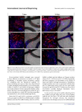
Figure 11. Immunofluorescence (red: F-actin; blue: nuclei) and bright-field images of cells and scaffolds after culture. HCFs on PCL/GelMA scaffolds with
two layers seven days after HCF culture: (A) PCL/GelMA_5, (B) PCL/GelMA_7, and (C) PCL/GelMA_10. HCFs on PCL/GelMA scaffolds with two layers
14 days after HCF culture: (D) PCL/GelMA_5, (E) PCL/GelMA_7, and (F) PCL/GelMA_10. White arrows denote cell colonization in the PCL area. Scale
bar: 100 µm. Abbreviations: HCF: Human cardiac fibroblasts; PCL/GelMA: Poly(ε-caprolactone)-gelatin methacryloyl.
Photocrosslinked GelMA hydrogels were prepared GelMA scaffolds reported stiffness and Young’s modulus
at three different concentrations via visible or UV light values of 0.51–1.18 N/mm and 6.8–10.5 MPa, respectively,
crosslinking to determine optimal biomimetic support and these parameters were dependent on the number of
for embedding cells into bioartificial scaffolds. UV layers and GelMA hydrogel concentration. Nonetheless,
photocrosslinking and exposure time were selected based Young’s modulus values were in agreement with literature
on photo-rheological characterization and Live/Dead assay. data on pathological cardiac tissue for men and women
GelMA_5, GelMA_7, and GelMA_10 hydrogels exhibited between the ages of 61 and 70. Furthermore, bioartificial
27
biomimetic stiffness (1–8 kPa) comparable to healthy cardiac PCL/GelMA scaffolds displayed biomimetic elastic
tissues. Moreover, sterilized hydrogels were stable for up to deformation of up to 22% strain. 25
14 days of incubation in sterile PBS. Their biocompatibility In vitro cell tests were performed by embedding HCFs
was assessed by in vitro cell tests on hydrogel eluates.
into GelMA hydrogels, filling the pores of PCL/GelMA
Before the PCL and GelMA combination, polyDOPA scaffolds with two layers. The cells were well-distributed
coating was applied on the PCL scaffolds to improve GelMA throughout the hydrogel. Likewise, the cytoskeletal
adhesion onto the PCL scaffold surface. Bioartificial PCL/ conformation was strongly influenced by GelMA hydrogel
Volume 10 Issue 3 (2024) 484 doi: 10.36922/ijb.2247