
Page 82 - IJB-4-2
P. 82
A multi-scale porous scaffold fabricated by a combined additive manufacturing and chemical etching process for bone tissue engineering
time point were used for the weight. Finally, the surface roughened surface feature with the extension of etching
morphologies and element distributions of the scaffolds time. This was attributed to the non-homogeneous
at different time points were investigated by FE-SEM hydrolysis of PLLA caused by the non-uniform local
and energy dispersive spectroscopy (EDS, X-Max20, crystallinity and cross-linking on the scaffolds surface.
Oxford Inc., UK). Subtle morphological variations were revealed at higher-
magnification images. It was found that the chemical
2.4 Statistical analysis etching process introduced micro pores (pits and
All quantitative results were expressed as means ± protrusions) throughout the struts of the scaffolds due
standard deviations. Statistical analysis was performed to the cleavage of ester bonds. A short etching time of
between different groups by Student’s t-test using SPSS 0.5 h produced some surface pores with characteristic
software (SPSS Inc., Chicago, IL, USA). And p values < sizes of 1~2 μm. As the etching time increased to 1.0 h,
0.05 were considered statistically significant. more PLLA was hydrolyzed, leaving well-ordered pore
arrangement on the scaffold surface with pore size
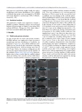
3. Results ranging from 1 μm to 3 μm. Moreover, these pores were
penetrated by smaller pores with pore size less than
3.1 Multi-scale porous structure 1 μm. In comparison, extending etching time to 1.5 h
To gain insight into the multi-scale porous structure, led to numerous pores with a range of sizes ranging
FE-SEM analysis was employed to study the porous from 1 μm to 10 μm on the struts. Such an increase
morphology and size distribution of scaffolds. As of surface roughness demonstrated a time-dependent
presented in Figure 2(a1), the scaffolds exhibited a well- etching degree and higher hydrophilicity compared to
defined porous network in three dimensions comprising PLLA-0 scaffold, facilitating the adhesion and ingrowth
interconnected pores, with an average pore size of of cells [30] . However, many cracks appeared on the
948±74 μm. Clearly, the chemical etching process did scaffold surface after 1.5 h of etching, which implied
not alter the original interconnected porous structure of that excessive etching might damage the original surface
the scaffolds (Figures 2(a2), 2(a3) and 2(a4)). Moreover, porous network or even the structure of bulk scaffold.
further observations revealed that the scaffold without These results confirmed that the multi-scale porous
chemical etching had a smooth surface while those structure could be fabricated and regulated by altering
chemically treated scaffolds exhibited a progressively the parameters of AM and chemical etching.
Figure 2. FE-SEM characterization of the multi-scale porous scaffolds: (a) interconnected porous network by AM, (b) Low- and (c) high-
magnification images of porous surface structure by chemical etching for different etching time.
4 International Journal of Bioprinting (2018)–Volume 4, Issue 2