
Page 147 - IJB-10-4
P. 147
International Journal of Bioprinting 3D printing innovations against infection
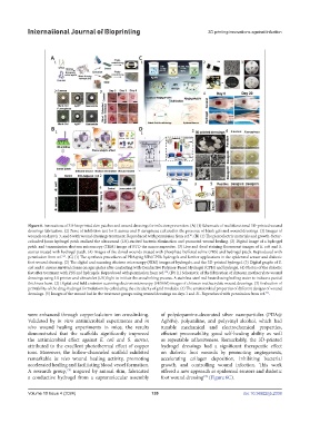
Figure 6. Innovations of 3D-bioprinted skin patches and wound dressings for infection prevention. (A) (1) Schematic of multifunctional 3D-printed wound
dressings fabrication. (2) Zone of inhibition test for S. aureus and P. aeruginosa cultured in the presence of blank gels and wound dressings. (3) Images of
wounds on days 0, 3, and 6 with wound dressings treatment. Reproduced with permission from ref. . (B) (1) The piezoelectric materials and growth-factor-
81
coloaded Janus hydrogel patch realized the ultrasound (US)-excited bacteria elimination and promoted wound healing. (2) Digital image of a hydrogel
patch and transmission electron microscopy (TEM) image of BTO-Au nanocomposites. (3) Live and dead staining fluorescent images of E. coli and S.
aureus treated with hydrogel patch. (4) Images of the dorsal wounds treated with phosphate-buffered saline (PBS) and hydrogel patch. Reproduced with
permission from ref. . (C) (1) The synthesis procedures of PDA@Ag NPs/CPHs hydrogels and further applications in the epidermal sensor and diabetic
169
foot wound dressing. (2) The digital and scanning electron microscopy (SEM) images of hydrogels, and the 3D-printed hydrogel. (3) Digital graphs of E.
coli and S. aureus survival clones on agar plates after contacting with Conductive Polymer-Based Hydrogel (CPH) and hydrogel. (4) Photos of the diabetic
feet after treatment with PBS and hydrogels. Reproduced with permission from ref. . (D) (1) Schematic of the fabrication of chitosan methacrylate wound
170
dressings using 3D printer and ultraviolet (UV) light to initiate the crosslinking process. A stainless-steel rod heated using boiling water to induce a partial
thickness burn. (2) Digital and field emission scanning electron microscopy (FESEM) images of chitosan methacrylate wound dressings. (3) Evaluation of
printability of the drug-hydrogel formulations by calculating the circularity of grid modules. (4) The antimicrobial properties of different designs of wound
dressings. (5) Images of the wound bed in the treatment groups using wound dressings on days 1 and 21. Reproduced with permission from ref. .
174
were enhanced through copper/calcium ion crosslinking. of polydopamine-decorated silver nanoparticles (PDA@
Validated by in vitro antimicrobial experiments and in AgNPs), polyaniline, and polyvinyl alcohol, which had
vivo wound healing experiments in mice, the results tunable mechanical and electrochemical properties,
demonstrated that the scaffolds significantly improved efficient processability, good self-healing ability as well
the antimicrobial effect against E. coli and S. aureus, as repeatable adhesiveness. Remarkably, the 3D-printed
attributed to the excellent photothermal effect of copper hydrogel dressings had a significant therapeutic effect
ions. Moreover, the hollow-channeled scaffold exhibited on diabetic foot wounds by promoting angiogenesis,
remarkable in vivo wound healing activity, promoting accelerating collagen deposition, inhibiting bacterial
accelerated healing and facilitating blood vessel formation. growth, and controlling wound infection. This work
A research group, inspired by animal skin, fabricated offered a new approach as epidermal sensors and diabetic
170
a conductive hydrogel from a supramolecular assembly foot wound dressing (Figure 6C).
170
Volume 10 Issue 4 (2024) 139 doi: 10.36922/ijb.2338