
Page 146 - IJB-10-4
P. 146
International Journal of Bioprinting 3D printing innovations against infection
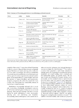
Table 3. Summary of 3D printing applications in wound dressings and dental materials
Device Author Benefits Materials 3D printer Ref.
Epidermal keratinocytes and
Albanna et al. Easy to access, precise positioning 3D inkjet 166
dermal fibroblasts
Enhances wound healing activity Hydrogel Alg-DA and gelatin
Chen et al. 3D inkjet 171
and blood vessel formation mixtures
On-demand printing of wound- Multilayer cell-hydrogel 3D free form
Lee et al. 175
Wound dressings specific skin layers composites fabrication
Promotes cell migration,
Cubo et al. proliferation, and differentiation in Human plasma-derived skin 3D free form 176
structure
fabrication
wound beds
Eliminates acoustic power bacteria New Janus piezoelectric hydrogel
Huang et al. 3D Extrusion 169
and promotes wound healing patch
Improves hydrophobicity and Chlorhexidine-loaded PDMS
Mai et al. DLP 179
antimicrobial activity of polymers based coated dental polymers
High mechanical strength and on- Dental fillings containing
Yang et al. 3D extrusion 180
demand drug release tinidazole
Selective laser
Sa et al. Sustains antimicrobial effect SLA resin containing Ag-HNT 181
curing
Dental material
Deng et al. Improves positioning accuracy and PLA and photopolymerization FDM and SLA 182
denture stability resins
Good workability and high
Sonaye et al. PEEK FFF 183
strength properties
Higher stiffness and elasticity as Resin with graphene nanosheets
Aati et al. DLP 184
well as biocompatibility added
Abbreviations: Ag-HNT, silver silicate nanotube; Alg, alginate; DA, dopamine; DLP, digital light processing; FDM, fused deposition modeling; FFF, fused
filament fabrication; PDMS, polydimethylsiloxane; PEEK, polyether ether ketone; PLA, polylactic acid; SLA, stereolithography.
a study by Albanna et al., researchers utilized 3D printing other antimicrobial substances, into hydrogel dressings is
166
to directly construct bilayered skin structures comprising common to mitigate the risk of infection. 167-170 For instance,
human fibroblasts and keratin-forming cells on defective a recent study generated a novel Janus piezoelectric
skin from nude mice and pigs. The outcomes revealed that hydrogel patch via 3D printing for sonodynamic bacteria
3D-printed materials expedited the wound healing process elimination and wound healing, which comprised gold
within a span of 3 weeks compared to the untreated control NP, methacrylate gelatin, and growth factors. The top
group. Immunohistochemical analysis further confirmed layer of patch can substantially eliminate infection under
the presence of human fibroblasts and keratinocytes, ultrasound, and the sustained release of growth factors
alongside endogenous cells, within the wound after 3 and from the bottom layer can promote tissue regeneration
169
6 weeks. This substantiates the considerable potential of during wound management (Figure 6B). The application
bioprinting in wound treatment. of 3D printing technology facilitates the even distribution
of these antimicrobial substances within the wound
Furthermore, biocompatible hydrogels play a crucial dressings, ensuring prolonged antimicrobial effects. In
role in providing a hydrophilic environment conducive a study conducted by Chen et al., coaxial 3D printing
to cell survival and have gained widespread use as wound technology was employed to successfully fabricate a hollow-
dressings. These hydrogel dressings contribute to wound channeled hydrogel scaffold, which comprises a mixture
healing by gently maintaining a moist environment, of dopamine-modified alginate (Alg-DA) and gelatin,
reducing pain, and minimizing scar formation. designed for treating bacterial infections in wounds. The
171
Incorporating antimicrobial agents, such as metal ions or scaffold’s structural stability and mechanical properties
Volume 10 Issue 4 (2024) 138 doi: 10.36922/ijb.2338