
Page 149 - IJB-10-4
P. 149
International Journal of Bioprinting 3D printing innovations against infection
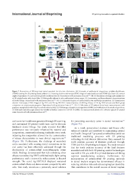
Figure 7. Innovations of 3D-bioprinted dental materials for infection elimination. (A) Schematic of antibacterial drug-release polydimethylsiloxane
(PDMS) coating for 3D printing dental polymer (1). Scanning electron microscopy (SEM) images of coated and uncoated PDMS specimens (2), contact
179
angles of specimens (3), and bacterial growth inhibition tests (4). Reproduced with permission from ref. . (B) (1) Illustration of design and manufacture
of 3D-printed molar with dental filler. (2) Biologically relevant in vitro antibacterial release assays. (3) Compression and biomechanical characterizations
180
of dental fillings. Reproduced with permission from ref. . (C) (1) Schematic diagram of antibacterial crown and bridge preparation. (2) Transmission
electron microscopy (TEM) images of Ag-HNT, and 3% Ag-HNT/SLR nanocomposites. (3) Killing efficacy of 1% Ag-HNT amount and blank group
181
composites on streptococcus pyogenes. Reproduced with permission from ref. . (D) (1) Schematic of 3D-printed dental resin nanocomposite with
graphene nanoplatelets with drug-free antimicrobial activity. (2) TEM images of graphene nanoparticles (GNPs) and verification of chemical compositions.
(3) SEM and confocal images present the viability of biofilm (control vs. 0.25 wt. % GNPs). (4) Antimicrobial activity of C. albicans against 3D-printed
resin. Reproduced with permission from ref. .
184
carious molar models were generated through 3D scanning, for preventing secondary caries in dental restorations
181
and customized 3D-printed molds were used to fabricate (Figure 7C).
tinidazole dental fillings. The study revealed that filler As a result, personalized dentures and braces offer
performance was intricately influenced by material and enhanced comfort and contribute to maintaining optimal
composition, consistently releasing tinidazole over a week. oral health. Deng et al. proposed a method that combines
182
Adjusting the composition allowed for the customization traditional machining processes with 3D printing
of release characteristics to meet clinical requirements technology for full denture fabrication. They assessed the
180
(Figure 7B). Furthermore, the challenge of secondary tooth position accuracy of dentures produced through
caries associated with wearing dental restorations in the FDM and SLA 3D printing techniques. The study revealed
oral cavity has been effectively addressed through the that the tooth position accuracy of the final dentures
development of antimicrobial stereolithography resins manufactured with both 3D printing-assisted technologies
(SLR). Findings demonstrated that SLR containing silver was within 150 μm, with SLA-printed dentures exhibiting
silicate nanotubes (Ag-HNT) exhibited stable light-curing superior manufacturing accuracy. Furthermore, the
performance and a noteworthy enhancement in flexural incorporation of antimicrobial 3D printing materials
strength. The cured Ag-HNT/SLR displayed sustained in dental implant surgery has demonstrated efficacy in
antimicrobial effects and demonstrated compatibility with reducing infection risks and enhancing implant durability.
cells. This study introduces a potentially novel solution This contributes to the overall success of dental implant
Volume 10 Issue 4 (2024) 141 doi: 10.36922/ijb.2338