
Page 174 - IJB-10-4
P. 174
International Journal of Bioprinting Printing collagen type IV membrane
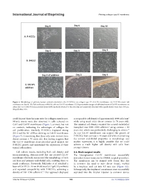
Figure 4. Morphology of primary human corneal endothelial cells (P-HCECs) on collagen type IV (Col-IV) membranes. (A) P-HCECs reach full
confluence on day 10. (B) Full confluence of B4G12 cells on Col-IV membrane. (C) Representative images of self-detachment of Col-IV membranes on
either day 3 or 5 with 5% human platelet lysate (hPL) (partially attached to the coverslip and completely detached with a gentle shake). Scale bars: 100 µm.
Magnification: 10×.
could deposit their laminin onto the collagen membranes. a comparable cell density of approximately 1000 cells/mm
2
Mitotic events were also observed in cells cultured on while using much older donor corneas (≥ 70 years old).
Col-I and Col-IV membranes (Figure 5, arrows), but not The minimal cell density required for corneal endothelial
2
on controls, indicating the advantages of collagen for transplant was 2000–2200 cells/mm using corneas < 30
cell proliferation. Similarly, P-HCECs displayed strong years old, which was particularly challenging to obtain. 34
ZO-1 and Na /K -ATPase staining on Col-IV membranes As our Col-IV membranes can support the growth of
+
+
(Figure 5). Considering that these cells were derived from P-HCECs from corneas ≥ 70 years old while maintaining
donor corneas ≥ 70 years old, this finding suggests that the correct endothelial properties in morphology and
the Col-IV membrane alone provided good support for marker expression, it is highly possible that we could
P-HCEC growth and maintained the expression of their achieve a much higher cell density with cells from
typical cell markers. younger donors.
Cell culture results, including both cell density and 3.5. Mock surgical results
immunostaining, demonstrated that our printed Col-IV The bioengineered Col-IV membranes successfully
membrane effectively maintained the morphology of both mimicked donor tissues in the DMEK surgical procedure.
cell lines and primary endothelial cells, enabling them to The membranes can be stained with vision blue dye
reach confluence. Previously, Palchesko et al. attached a (a common dye used to stain donor tissue), marked
layer of Col-IV (5–10 nm thick) to a Col-I gel (10 µm thick) by a trephine, and cut into 8.5 mm size (Figure 6A).
and reported compatibility with P-HCECs, achieving a Subsequently, the trephined membranes were successfully
density of 760–1700 cells/mm . Our approach displayed aspirated into the Stryker injector (a common device
2 11
Volume 10 Issue 4 (2024) 166 doi: 10.36922/ijb.3258