
Page 175 - IJB-10-4
P. 175
International Journal of Bioprinting Printing collagen type IV membrane
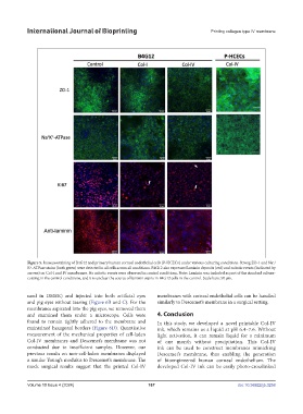
Figure 5. Immunostaining of B4G12 and primary human corneal endothelial cells (P-HCECs) under various culturing conditions. Strong ZO-1 and Na /
+
K -ATPase stains (both green) were detected in all cells across all conditions. B4G12 also expressed laminin deposits (red) and mitotic events (indicated by
+
arrows) on Col-I and IV membranes. No mitotic events were observed in control conditions. Note: Laminin was included as part of the standard culture-
coating in the control conditions, and it is unclear the source of laminin stains in B4G12 cells in the control. Scale bars: 50 µm.
used in DMEK) and injected into both artificial eyes membranes with corneal endothelial cells can be handled
and pig eyes without tearing (Figure 6B and C). For the similarly to Descemet’s membrane in a surgical setting.
membranes aspirated into the pig eyes, we removed them
and examined them under a microscope. Cells were 4. Conclusion
found to remain tightly adhered to the membrane and In this study, we developed a novel printable Col-IV
maintained hexagonal borders (Figure 6D). Quantitative ink, which remains as a liquid at pH 6.4–7.6. Without
measurement of the mechanical properties of cell-laden light activation, it can remain liquid for a minimum
Col-IV membranes and Descemet’s membrane was not of one month without precipitation. This Col-IV
conducted due to insufficient samples. However, our ink can be used to construct membranes mimicking
previous results on non-cell-laden membranes displayed Descemet’s membrane, thus enabling the generation
a similar Young’s modulus to Descemet’s membrane. The of bioengineered human corneal endothelium. The
mock surgical results suggest that the printed Col-IV developed Col-IV ink can be easily photo-crosslinked
Volume 10 Issue 4 (2024) 167 doi: 10.36922/ijb.3258