
Page 176 - IJB-10-4
P. 176
International Journal of Bioprinting Printing collagen type IV membrane
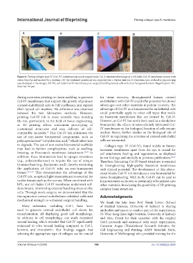
Figure 6. Testing collagen type IV (Col-IV) membranes in mock surgical trials. (A) A representative image of a cell-laden Col-IV membrane stained with
vision blue dye and marked by a trephine. (B) The trephined membrane was aspirated into a Stryker injector. (C) Injection into artificial or pig eyes (pig
eyes displayed in the image). (D) The cell-laden Col-IV membrane post-surgical handling reveals cells with clear hexagonal borders. Magnification: 10×.
Scale bar: 50 µm.
during extrusion printing or classic molding to generate for tissue recovery. Bioengineered human corneal
Col-IV membranes that support the growth of primary endothelium with Col-IV could offer potential functional
corneal endothelial cells to full confluence and express advantages over other materials in patient recovery. The
their typical cell markers. No difference was observed advantage of Col-IV as a biomaterial for endothelial cells
between the two fabrication methods. However, could potentially apply to other cell types that reside
printing Col-IV ink is more versatile than molding on basement membranes that are formed by Col-IV.
the ink, particularly in the field of tissue engineering, However, as Col-IV has rarely been used as a standalone
as 3D printing allows convenient prototyping of biomaterial, the effects of extracellularly fabricated Col-
customized structures and easy delivery of cell- IV membranes on the biological function of cells remain
compatible materials. This Col-IV ink eliminates the unclear. Hence, further studies on the biological role of
35
use of non-native biomaterial components, such as Col-IV in regulating the activities of corneal endothelial
37
36
polycaprolactone or hyaluronic acid, which takes time cells are warranted.
to degrade. The use of non-native biomaterial scaffolds Collagen type IV (Col-IV), found widely in human
may lead to further complications, such as swelling, basement membranes apart from the eye, is crucial for
bruising, or Descemet’s membrane detachment. 38,39 In cell attachment, healing, and regeneration, as displayed
addition, these biomaterials lead to opaque structures in our findings and similarly in previous publications. 4,5,27
(e.g., polycaprolactone) or require the use of opaque Therefore, fabricating Col-IV-based structures is essential
titanium base (e.g., hyaluronic acid), thereby restricting in bioengineering high-quality basement membranes
the application of Col-IV inks on non-transparent with clinical potential. The development of this photo-
tissues. 36,37,40 This demonstrates the advantage of the crosslinkable Col-IV ink introduces a new biomaterial for
Col-IV ink, as optical light transmittance is essential for tissue bioengineering. With its Pr, Col-IV can be used to
ocular tissues such as the cornea. When combined with form structures on its own or potentially to be printed onto
hPL, our cell-laden Col-IV membrane underwent self- other materials, broadening the possibility of 3D printing
detachment, minimizing external handling stress on the complex tissue structures.
cells. Through mock surgery, we demonstrated that the
bioengineered corneal endothelium exhibited sufficient Acknowledgments
mechanical strength to withstand surgical handling.
We thank the help from Prof. Frank Lovicu (School
Many substrates, including Col-I, have been of Medical Sciences, University of Sydney) in sharing
used to generate corneal endothelial cell sheets for antibodies and spaces to conduct part of the experiments,
transplantation, all displaying good cell morphology. Dr Yihui Song (Save Sight Institute, University of Sydney)
In addition to cell morphology, our study examined and Arzu Demir for their expertise with the original
wound healing, which revealed that Col-IV significantly Col-I protocols and assistance with some experiments,
enhanced in vitro wound healing compared to Col-I, Cameron Angus (Translational Research Initiative for
laminin, and chondroitin. Our findings suggest that Cell Engineering and Printing, ANFF Materials Node,
selecting the appropriate type of collagen can be crucial University of Wollongong) who provided training for the
Volume 10 Issue 4 (2024) 168 doi: 10.36922/ijb.3258